





Macrophages play a crucial role in defending against microbial infections, but their abnormal activation can trigger inflammation and lead to significant histopathological changes. Notable histopathological alterations are observed in atherosclerotic plaques, and abnormal macrophage activation is also present in autoimmune diseases such as rheumatoid arthritis and inflammatory bowel disease. Rheumatoid arthritis is characterized by macrophage and lymphocyte infiltration into the synovium, with macrophage quantity and activation state correlating with disease progression. Macrophage activation is determined by a series of signals, including cytokines and microbial molecules. Interferon-γ is a classical M1 macrophage activation factor that enhances lipopolysaccharide-stimulated macrophage activation. It remains unclear whether receptors regulating macrophage hyperactivation exist in human inflammatory diseases. FITC-labeled SLAMF7/CRACC/CD319 Fc chimera protein can be used to quantitatively detect SLAMF7 protein expression levels and binding activity, providing a technical tool for inflammatory disease research.
Researchers used rheumatoid arthritis as a model to identify receptors for macrophage hyperactivation. RNA-seq analysis revealed that, compared to non-inflammatory osteoarthritis, rheumatoid arthritis patients exhibited high expression of interferon-induced genes, inflammatory cytokines, and chemokines, indicating macrophage activation is associated with elevated interferon and inflammatory factors. Omics results showed SLAMF7 as the most differentially expressed gene. SLAMF7 is a member of the signaling lymphocytic activation molecule family 7, expressed as an activation receptor on natural killer cells and immune cell subsets. Isolation of synovial tissue macrophages from osteoarthritis and rheumatoid arthritis patients revealed that SLAMF7 expression was significantly higher in rheumatoid arthritis, with SLAMF7 detected in macrophages of up to 55% of rheumatoid arthritis patients. Consistent results were obtained in synovial fluid macrophages.
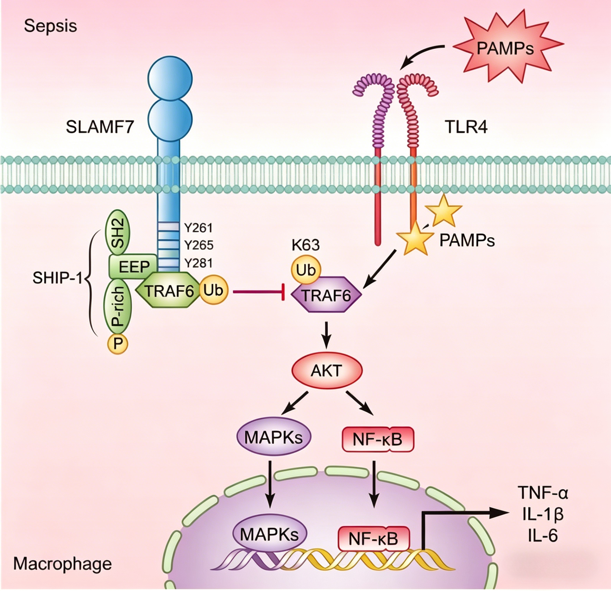
Under non-stimulated conditions, SLAMF7 is highly expressed in plasma cells but low in macrophages. However, high SLAMF7 expression has been observed in macrophages from atherosclerotic lesions and bone marrow fibrosis patients. Differential gene expression analysis of SLAMF7-high and SLAMF7-low cells isolated from CD14-positive and CD16-positive peripheral blood cells of healthy individuals or rheumatoid arthritis patients revealed that high SLAMF7 expression is often accompanied by elevated interferon-induced genes. Gene enrichment analysis further confirmed that SLAMF7 high expression correlates with type I and II interferon responses. In vitro experiments showed that interferon stimulation significantly increased SLAMF7 expression in macrophages. Cytokines such as IFN-β, IL-1β, and TNF-α, as well as TLR agonists, could upregulate SLAMF7 expression on macrophages. Treatment with JAK1 and JAK2 inhibitors or siRNA targeting the interferon receptor γ significantly reduced SLAMF7 expression, confirming interferon-γ as a key regulator of SLAMF7 expression in macrophages.
Researchers investigated whether SLAMF7 activates or inhibits macrophages. Interferon-γ treatment increased TNF-α mRNA levels in macrophages, but SLAMF7 silencing did not further enhance TNF-α production, indicating SLAMF7 does not suppress macrophage activation. Pretreatment of macrophages with interferon-γ upregulated SLAMF7, and subsequent addition of recombinant SLAMF7 protein increased TNF-α mRNA by 80-fold. Conversely, SLAMF7 silencing significantly reduced TNF-α expression, demonstrating that SLAMF7 further enhances interferon-γ-induced macrophage activation. RNA sequencing revealed that SLAMF7 participates in dramatic changes in inflammatory factors (TNF, IL-1β, IL-6, and IL-12β) and chemokines, alongside downregulation of mitochondrial and ribosomal pathway genes, suggesting a metabolic shift that supports macrophage hyperactivation. FITC-labeled SLAMF7/CRACC/CD319 Fc chimera protein can be used to study SLAMF7 interactions with downstream signaling molecules and assess its role in enhancing macrophage activation.
Macrophages express two ITAM-containing proteins, DAP12 and FcGammaR, as well as EAT-2. High levels of FCER1G and TYROBP were detected in synovial macrophages, but SH2D1B was absent, indicating EAT-2 is not involved in macrophage activation. Silencing FCER1G reduced TNF-α production in macrophages stimulated with recombinant SLAMF7, suggesting FcGammaR plays a critical role in SLAMF7-mediated macrophage activation. Researchers found that SLAMF7 activation of macrophages significantly increased phosphorylation of ERK, NF-κB, MAPK, and AKT, activating multiple signaling pathways to reprogram macrophage metabolism and drive inflammatory gene expression. Additionally, TNF-α may amplify signaling via autocrine pathways, as TNF secretion was significantly reduced after TNFR silencing or anti-TNFR antibody treatment, implying TNF-α autocrine signaling contributes to macrophage hyperactivation.
SLAMF7 is significantly upregulated in inflammatory rheumatoid arthritis, inflammatory bowel disease, and COVID-19 pneumonia, reinforcing the role of hyperactivated macrophages in driving inflammation in autoimmune diseases and infections. Targeting this receptor may provide new clinical treatment avenues. FITC-labeled SLAMF7/CRACC/CD319 Fc chimera protein offers a sensitive and efficient detection tool for studying SLAMF7 protein expression, distribution, and interactions, applicable in macrophage function assessment and immune monitoring.
Nanjing UA-BioTech Co., Ltd. (UA-Bio) has independently developed "FITC-Labeled SLAMF7/CRACC/CD319 Fc Chimera Protein, Human" (Catalog No.: UA011262), a high-performance green fluorescent-labeled probe specifically designed for SLAMF7-targeted research and multiple myeloma immunotherapy evaluation. SLAMF7 (also known as CRACC or CD319), a member of the signaling lymphocytic activation molecule family, is highly expressed on the surface of multiple myeloma cells and is a critical target for tumor immunotherapy. This protein is a human SLAMF7 extracellular domain fused with human IgG Fc fragment and labeled with FITC fluorescence, enabling efficient binding to anti-SLAMF7 antibodies or affinity ligands. It serves as a stable and reliable standardized tool for CAR-T cell therapy evaluation, antibody drug screening, multiple myeloma biomarker analysis, and more.
| Core Advantages | Detailed Parameters / Functional Description |
|---|---|
| High Purity and Intact Bioactivity | The product utilizes an advanced eukaryotic expression system and highly standardized purification processes, validated by multi-dimensional quality control to ensure >95% purity and correct native conformation (with intact glycosylation modifications). The Fc chimera form enhances protein stability and proper folding. The optimized FITC labeling process ensures high labeling efficiency while maintaining high antibody-binding affinity, accurately simulating SLAMF7 antigenic properties on multiple myeloma cells.-- |
| High-Brightness FITC Labeling | An optimized FITC labeling ratio ensures stable fluorescent dye labeling per SLAMF7-Fc chimera protein molecule, producing bright green fluorescence signals under 488 nm excitation. Ideal for flow cytometry (FACS) and fluorescence microscopy, providing reliable support for SLAMF7-targeted molecule activity evaluation and precise quantification.-- |
| Exceptional Batch Consistency and Stability | Strict management from protein expression to labeling and purification, combined with comprehensive release testing, ensures consistent binding activity, uniform fluorescence intensity, and excellent long-term stability. Provides solid quality assurance for long-term SLAMF7-targeted research.-- |
| Versatile Application Tool | This protein excels in CAR-T cell positivity rate flow detection, anti-SLAMF7 antibody/antagonist screening (e.g., elotuzumab biosimilar activity evaluation), competitive binding assays, multiple myeloma biomarker analysis, and flow cytometry. Suitable for SLAMF7-targeted CAR-T cell therapy development, diagnostic tool research, and drug activity evaluation.-- |
| Comprehensive Solutions and Professional Support | We provide validated standard protocols, typical flow cytometry data, and detailed interpretation guides to help establish stable and reproducible SLAMF7-targeted molecule detection workflows. Nanjing UA-Bio's technical team offers professional consultation and support for research design, optimization, and data analysis.-- |
Nanjing UA-BioTech Co., Ltd. is committed to providing cutting-edge, high-quality core reagents and tools for immunology, cell therapy, and innovative drug development. For detailed technical parameters, validation data, or application inquiries regarding "FITC-Labeled SLAMF7/CRACC/CD319 Fc Chimera Protein, Human" (Catalog No.: UA011262), please feel free to contact us.








