




The cytokine regulatory mechanism and research applications of human monocyte differentiation into osteoclasts
Author: Senior Product Manager of UA Views:144 


Osteoclasts are the only multinucleated giant cells in the human body capable of bone resorption, and their differentiation and maturation are precisely regulated by various cytokines. Human peripheral blood CD14+ monocytes can be directionally differentiated into mature osteoclasts with bone resorption activity under the synergistic induction of macrophage colony-stimulating factor and receptor activator of nuclear factor kappa-B ligand. This in vitro induction system has become a core experimental platform for bone metabolism disease research, drug screening, and mechanism analysis.
- Recent Advances
- Product Information
Recent Advances
Cytokine Regulatory Mechanisms and Research Applications of Human Monocyte Differentiation into Osteoclasts
Brief Description: Osteoclasts are the only multinucleated giant cells in the human body capable of bone resorption, and their differentiation and maturation are precisely regulated by various cytokines. Human peripheral blood CD14+ monocytes can be directionally differentiated into mature osteoclasts with bone resorption activity under the synergistic induction of macrophage colony-stimulating factor (M-CSF) and receptor activator of nuclear factor kappa-B ligand (RANKL). This in vitro induction system has become a core experimental platform for bone metabolism disease research, drug screening, and mechanistic analysis.
Biological Origin of Osteoclasts and Their Central Role in Bone Homeostasis.
Osteoclasts are multinucleated giant cells of the hematopoietic lineage and are the only terminally differentiated cells in the skeletal system capable of performing bone resorption. Under physiological conditions, osteoclasts participate in bone development, fracture repair, and bone homeostasis maintenance through precise bone resorption activities. However, when osteoclast differentiation or activity becomes abnormal, the dynamic balance between bone resorption and bone formation is disrupted, leading to pathological bone loss diseases such as osteoporosis, rheumatoid arthritis bone destruction, and bone tumor metastasis.
During development, osteoclasts primarily originate from bone marrow erythroid progenitors, while osteoclasts required for maintaining bone homeostasis in adulthood are derived from circulating monocytes. Human peripheral blood CD14+ monocytes, as an important precursor source of osteoclasts, have a differentiation capacity closely related to the occurrence and progression of various bone metabolism diseases.
Core Cytokines Driving Osteoclast Differentiation and Their Signaling Mechanisms.
Macrophage colony-stimulating factor (M-CSF) and receptor activator of nuclear factor kappa-B ligand (RANKL) are the two key cytokines that induce monocyte differentiation into osteoclasts.
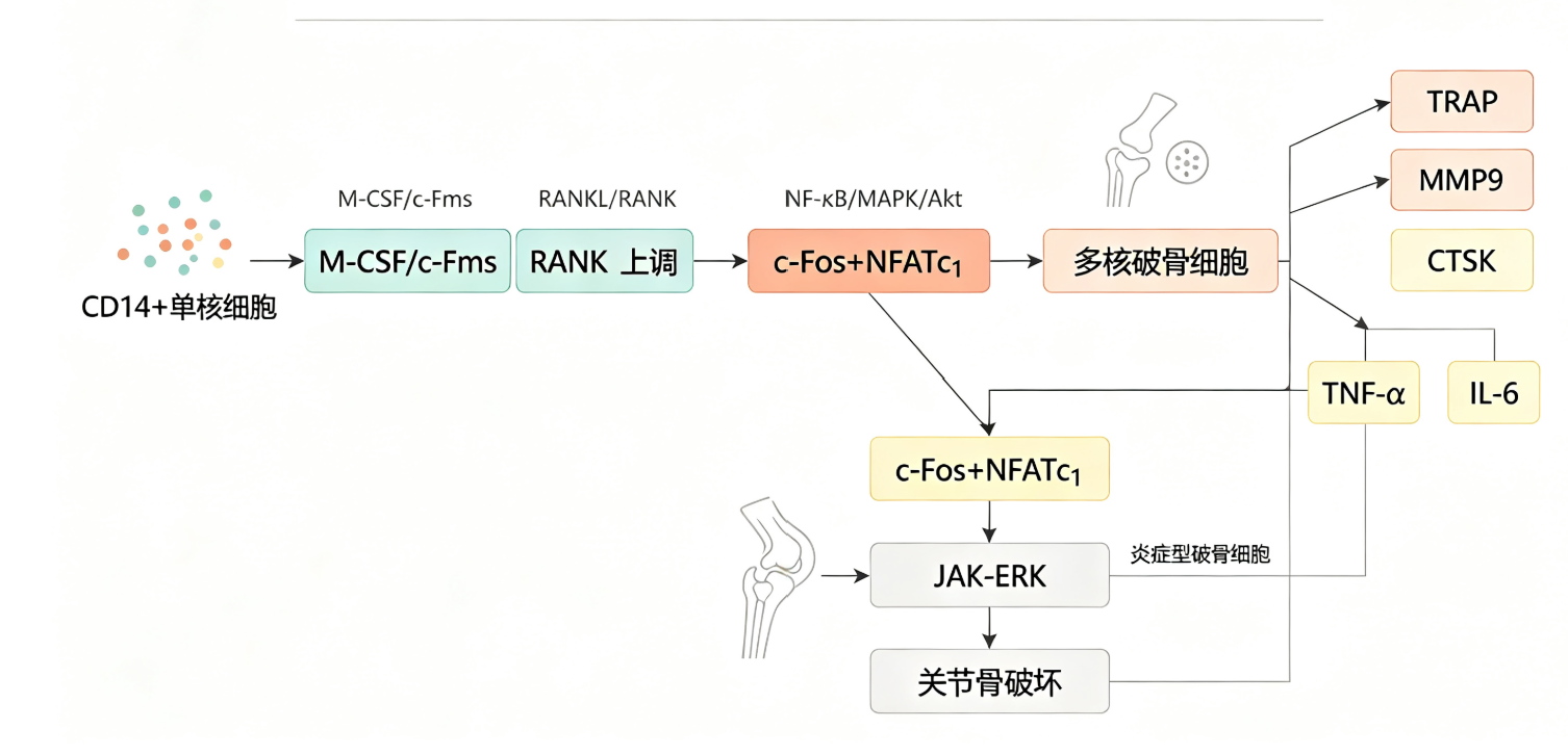
M-CSF binds to the c-Fms receptor on the monocyte surface, playing a fundamental role in promoting the survival, proliferation, and differentiation readiness of osteoclast precursor cells. Under M-CSF stimulation, monocytes upregulate the expression of RANK on their cell membranes, preparing them for subsequent RANKL signaling.
As a member of the tumor necrosis factor superfamily, RANKL binds to the RANK receptor on osteoclast precursor cells, activating multiple signaling pathways such as NF-κB, MAPK, ERK, and PI3K/Akt, ultimately inducing the expression of key transcription factors like c-Fos and NFATc1. These factors drive the programmed expression of osteoclast-specific genes, including tartrate-resistant acid phosphatase (TRAP), cathepsin K, and matrix metalloproteinase-9 (MMP-9), which collectively confer osteoclasts with their unique bone resorption capability. Under continuous RANKL stimulation, monocytes gradually fuse to form multinucleated giant cells, i.e., mature osteoclasts.
Complementary Regulatory Role of Inflammatory Cytokines in Osteoclast Differentiation.
In addition to the classical M-CSF and RANKL pathway, inflammatory cytokines such as tumor necrosis factor-alpha (TNF-α) and interleukin-6 (IL-6) can also directly induce osteoclast differentiation. In inflammatory diseases like rheumatoid arthritis, high levels of TNF-α and IL-6 in the joint microenvironment activate c-Fos and NFATc1 through the JAK-ERK signaling pathway, bypassing the RANKL-dependent pathway to directly drive osteoclast formation.
This inflammation-driven osteoclast differentiation pathway has significant pathophysiological implications. TNF-α and IL-6-induced osteoclasts highly express interleukin-1β (IL-1β), interleukin-12 (IL-12), and matrix metalloproteinase-3 (MMP-3), exhibiting biological characteristics distinct from RANKL-induced osteoclasts. Clinical studies have shown that peripheral blood monocytes from rheumatoid arthritis patients have a stronger osteoclast differentiation capacity, and the number of inflammation-induced osteoclasts is positively correlated with the progression of joint structural damage.
Standardized Experimental Protocol for In Vitro Osteoclast Differentiation.
Based on the above cytokine regulatory mechanisms, researchers have established a standardized protocol for in vitro differentiation of osteoclasts from human peripheral blood CD14+ monocytes. This protocol consists of three key steps.
First, peripheral blood mononuclear cells (PBMCs) are isolated from human peripheral blood via density gradient centrifugation, followed by CD14 magnetic bead positive selection to enrich CD14+ monocytes. Second, the purified CD14+ monocytes are seeded in culture medium containing M-CSF and cultured overnight to allow full adhesion and upregulation of RANK expression. Third, RANKL is added to the M-CSF-maintained culture for induction, and multinucleated osteoclasts can be obtained after 5 to 7 days of culture.
The identification of induced osteoclasts typically involves tartrate-resistant acid phosphatase (TRAP) staining and morphological observation. Mature osteoclasts exhibit a typical multinucleated giant cell morphology under optical microscopy, with red or purple-red cytoplasm after TRAP staining. To further verify the functional activity of osteoclasts, methods such as bone resorption pit assay, F-actin ring immunofluorescence staining, and ATP production measurement can be employed.
Application Value and Research Tools of the Osteoclast Differentiation System.
This in vitro induction system has become an important experimental platform in the field of bone metabolism research, widely used for mechanistic analysis of osteoclast differentiation, bone remodeling regulation studies, exploration of pathological mechanisms in bone metabolism diseases, and high-throughput screening of anti-resorptive drugs. Based on this system, researchers can evaluate the osteoclast differentiation potential of circulating monocytes under disease conditions and screen therapeutic compounds targeting osteoclast differentiation and function.
In terms of experimental tools, Nanjing YouAi offers a human monocyte-induced osteoclast differentiation cytokine kit, providing a standardized solution for this research field. The kit precisely includes recombinant human M-CSF and recombinant human RANKL, along with a complete protocol for isolation, induction, and identification. Validated by TRAP staining, the mature osteoclasts show specific positive reactions, making it suitable for studies on osteoclast differentiation mechanisms, bone remodeling regulation, and anti-resorptive drug screening.
This article is reviewed and published by the technical expert team of UA
Disclaimer: This article partially utilizes artificial intelligence assistance in its creation. If any content involves copyright or intellectual property issues, please let us know and we promise to verify and remove it as soon as possible.
Purchase recombinant protein, choose Nanjing UA-Bio
UA protein focuses on providing various protein reagents, raw materials, and services required for drug research and development, cell therapy, gene therapy, and basic scientific research, including drug target proteins, immune checkpoint proteins, cytokines, tool enzymes, customized protein expression, and full-length transmembrane protein development. Youai is committed to providing customers with high-quality products and professional services, and building a High-tech Biological Enterprise with International Competitiveness.
Target proteins | membrane proteins | cytokines | enzymes | viral antigens | protein customization
Nanjing UA Biotechnology Co., Ltd. Email:order@ua-bio.com Phone:+86-25-56221161

Product Information

Link:





©2021-2026 all rights reserved UA Biotechnology Co.,Ltd.
Privacy Policy 
网站备案号 浙ICP备2022019033号-2 浙公网安备33010202004867号
 Business license (three certificates in one)
 Tel:+86-25-56221161
E-mail:order@ua-bio.com
All products sold on this website shall not be used for clinical diagnosis or treatment of humans or animals, and shall only be used for non-medical purposes such as industrial or scientific research.
