




Caspase 3/7 Assay Kit Standard Operating Procedure and Result Interpretation


Accurate detection of apoptosis is a critical step in life science research, and the activation of Caspase 3/7 is a hallmark event marking the irreversible execution phase of apoptosis. This guide provides a detailed standard operating procedure for fluorescence-based Caspase 3/7 detection kits, helping you obtain reliable and reproducible experimental data.
- Recent Advances
- Product Information
Accurate detection of apoptosis is a critical step in life science research, and the activation of Caspase 3/7 is a hallmark event marking the irreversible execution phase of apoptosis. This guide will detail the standard operating procedures for fluorescence-based Caspase 3/7 detection kits to help you obtain reliable and reproducible experimental data.
I. Pre-experiment Preparation and Experimental Design
Successful detection begins with thorough experimental planning.
1. Cell Preparation and Plating
Plate Selection: Always use white 96-well or 384-well cell culture plates. The white walls effectively reflect and enhance weak fluorescent signals, improving detection sensitivity and signal-to-noise ratio compared to transparent standard plates.
Cell Density: Seeding cells at an "appropriate density" is crucial. Too low density results in weak signals undetectable; too high density may cause contact inhibition or nutrient depletion, generating nonspecific background. Optimize through preliminary experiments, typically seeding 5,000 - 50,000 cells per well in 96-well plates (varies by cell type).
2. Key Experimental Controls
Well-designed controls are the cornerstone of data interpretation. Be sure to include the following control wells:
Negative Control: Cells treated with solvent only (e.g., DMSO). This group establishes the baseline Caspase 3/7 activity level without apoptosis induction.
Blank Control: Medium-only wells without cells. Used to subtract background fluorescence from the medium itself.
Positive Control (Highly Recommended): Cells treated with known apoptosis inducers (e.g., staurosporine, camptothecin). Validates your detection system and reagent functionality while providing a reference signal intensity.
II. Detailed Detection Steps
Follow this protocol strictly to ensure consistent results.
Step 1: Apoptosis Induction and Incubation
After cells adhere and grow to an appropriate state, add your test compounds to designated wells.
Gentle Mixing: After addition, ensure uniform compound distribution by lightly shaking the plate or using a multichannel pipette (avoid bubbles).
Return cells to the incubator for a predetermined induction period (optimized via preliminary experiments).
Step 2: Reagent and Cell Equilibrium
Before detection, equilibrate both the Caspase 3/7 detection reagent and cell plate at room temperature for 20 minutes.
Critical: This step prevents temperature shock (cold reagent → 37°C cells) that may affect enzyme activity or cause condensation, compromising readout accuracy. Room-temperature reactions yield more stable results.
Step 3: Homogeneous Detection — Adding Detection Reagent
Add detection reagent at recommended volumes:
96-well plate: Add 50 µL reagent to 100 µL medium.
384-well plate: Add 10 µL reagent to 20 µL medium.
Mixing and Incubation:
Mix thoroughly using a plate shaker for 2 minutes.
Incubate in darkness at room temperature for 30-60 minutes.
Light Protection: Prevents fluorophore quenching.
Time Window: Fluorescence typically peaks ~1 hour post-reagent addition. Early reads at 30 minutes are possible, but optimal signals occur near 60 minutes. Avoid exceeding 3 hours to prevent signal decay or elevated background.
Step 4: Signal Measurement
Use a chemiluminescence-capable microplate reader with correct parameters (often broadband detection without filters).
Read immediately, maintaining consistent intervals between wells for comparable data.
III. Principle Review and Data Analysis
1. Working Principle
The kit employs cell-permeable fluorescent substrates (e.g., DEVD peptide-linked fluorophores). During apoptosis, activated Caspase 3/7 cleaves the substrate, releasing fluorophores to generate fluorescence proportional to enzyme activity (i.e., apoptotic cell number/degree).
2. Data Analysis
Data Export: Transfer raw fluorescence values (RLU) to analysis software.
Background Subtraction: Subtract "blank control" averages from all wells.
Calculations:
Direct comparison: Compare background-subtracted fluorescence across treatment groups.
Normalization: Express treatment signals relative to "negative control" means as fold induction (treatment signal/control signal).
Statistics: Apply t-tests or ANOVA to assess significance between groups.
Conclusion
Following this SOP enables reliable assessment of compound-induced apoptosis using Caspase 3/7 detection kits. Rigorous design, standardized protocols, and rational analysis are key to unraveling molecular mechanisms governing cell fate decisions.
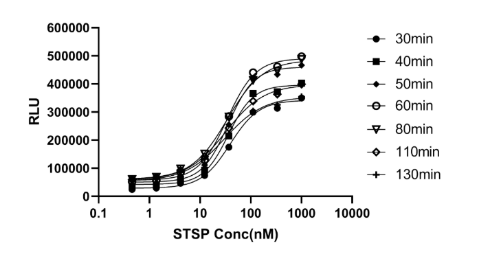
Caspase 3/7 apoptosis assay evaluating staurosporine (STSP) effects on HeLa cells: HeLa cells (4×10⁵/mL) were seeded in white 96-well clear-bottom plates (100 µL/well). After 24 hours, 3-fold serial diluted STSP was added, followed by 6-hour incubation (37°C, 5% CO2). Caspase 3/7 activity was measured per kit instructions. Figure 1 shows dose-response curves (fluorescence at 30-130 minutes), with tabulated EC50 values.
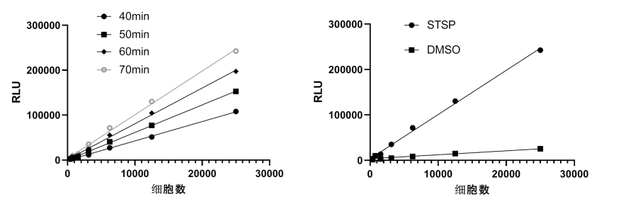
Caspase 3/7 assay sensitivity test: HeLa cells treated with 1 µM STSP for 6 hours were serially diluted two-fold and seeded (25,000–180 cells/well) in 96-well clear-bottom white plates (100 µL/well). Caspase 3/7 activity was measured per protocol. Left panel: Fluorescence at 40-70 minutes showed linear correlation (R2>0.99) with apoptotic cell numbers, detecting as few as 200 apoptotic cells. Right panel: Fluorescence (70 minutes) from HeLa cells treated with 1 µM STSP vs. DMSO solvent control.
This article is reviewed and published by the technical expert team of UA
Disclaimer: This article partially utilizes artificial intelligence assistance in its creation. If any content involves copyright or intellectual property issues, please let us know and we promise to verify and remove it as soon as possible.
Purchase recombinant protein, choose Nanjing UA-Bio
UA protein focuses on providing various protein reagents, raw materials, and services required for drug research and development, cell therapy, gene therapy, and basic scientific research, including drug target proteins, immune checkpoint proteins, cytokines, tool enzymes, customized protein expression, and full-length transmembrane protein development. Youai is committed to providing customers with high-quality products and professional services, and building a High-tech Biological Enterprise with International Competitiveness.







 Business license (three certificates in one)