




Luminescent vs. Fluorescent: How to Choose a Cell Viability Assay?


In the fields of life sciences and drug development, accurately, rapidly, and high-throughput assessing cell viability is a fundamental and critical step. Whether evaluating compound toxicity, screening anticancer drugs, or optimizing cell culture conditions, reliable cell viability detection technologies are essential.
- Recent Advances
- Product Information
In the fields of life sciences and drug development, accurately, rapidly, and high-throughput assessing cell viability is a fundamental and critical step. Whether evaluating compound toxicity, screening anticancer drugs, or optimizing cell culture conditions, reliable cell viability detection technologies are essential. New-generation detection kits centered on luminescent and fluorescent methods are leading this field toward greater precision and efficiency. This article will highlight several representative advanced detection solutions.
I. The Gold Standard: Classic Luminescent Cell Viability Assay
Core Principle:
Based on the ATP-dependent luciferase-catalyzed reaction. In the presence of Mg²⁺, luciferase uses luciferin, ATP, and O₂ as substrates to convert chemical energy into light energy. Since ATP is the core molecule of energy metabolism in living cells, its content is linearly positively correlated with the number of viable cells. Therefore, luminescence intensity directly reflects cell viability.
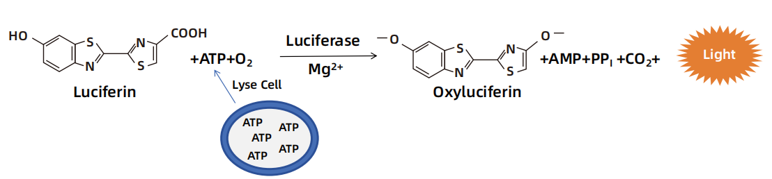
Technical Features:
High Sensitivity and Wide Dynamic Range:
Can detect very small numbers of cells and cover a wide range of cell concentrations.
Simple Operation and Homogeneous Detection:
"Add-incubate-read" in one step, no washing required, especially suitable for high-throughput screening (HTS).
Low Background:
Bioluminescence background is extremely low, with a high signal-to-noise ratio.
Typical Applications:
Cell proliferation/toxicity screening, drug sensitivity testing, antibody-dependent cellular cytotoxicity (ADCC) effect evaluation, etc.
| Catalog No. | Product Name |
|---|---|
| UA070103 | UA-Glo® Luminescent Cell Viability Assay |
II. Performance Upgrade: Luminescent Cell Viability 2.0 Assay
As an upgraded version of the classic ATP assay, the 2.0 Assay has been optimized in multiple dimensions:
1. Enhanced Signal Stability: The improved reagent formulation allows luminescence signals to last for hours or even longer, alleviating the stringent timing requirements for reading in high-throughput screening and enabling simultaneous processing of multiple plates.
2. Higher Sensitivity and Wider Linear Range: Can detect lower numbers of cells and maintain a linear response to higher cell concentrations, suitable for more diverse cell types and experimental conditions.
3. Optimized Interference Resistance: Greater tolerance to common components in culture media (e.g., phenol red, serum, drug compounds), reducing false-negative or false-positive results.
4. Improved Reagent Stability: Reagents remain active for longer periods, facilitating ease of use.
Selection Guide:
When conducting large-scale compound library screening or using special cell lines sensitive to detection conditions, the 2.0 version provides more stable and reliable data quality.
| Catalog No. | Product Name |
|---|---|
| UA079014 | UA-Glo® Luminescent Cell Viability 2.0 Assay |
III. Breaking the Bottleneck of 3D Culture Detection: 3D Cell Viability Assay
Three-dimensional cell cultures (e.g., spheroids, organoids) can more realistically simulate the in vivo microenvironment, but their dense structures pose new challenges for reagent penetration.
Core Challenges and Solutions:
Traditional detection reagents struggle to penetrate the interior of 3D spheroids effectively, leading to signal distortion. The dedicated 3D Cell Viability Assay addresses this through:
Powerful Lysis and Penetration Reagents: Ensure complete lysis of all cells inside and outside the spheroid, releasing intact ATP.
Combination with ATP Detection Technology: Typically paired with the highly sensitive luminescent method (ATP detection), total ATP content is measured after thorough lysis to reflect the cell viability of the entire 3D structure.
Standardized Workflow: Provides optimized protocols for 3D spheroid preparation, processing, lysis, and detection.
Typical Applications:
Evaluation of antitumor drug efficacy and penetration in solid tumor models, developmental toxicity testing in stem cell organoids, more realistic disease modeling, and assessment of compound toxicity and efficacy in 3D organoids.
| Catalog No. | Product Name |
|---|---|
| UA079011 | UA-Glo® 3D Cell Viability Assay |
IV. Multiparametric Fluorescent Detection: Fluorescent Cell Viability Assay
Fluorescent methods use different fluorescent probes to label cell structures or metabolic activity, enabling multidimensional viability assessment.
Common Types and Principles:
Based on the degradation of peptide-fluorophore substrates by proteases present only in living cells, which generates fluorescent signals. When cell membrane integrity is compromised, these proteases lose activity and no fluorescence is produced. Unlike our Luminescent Cell Viability Assay (Catalog No. UA070103), this method can detect earlier and more subtle cell damage, and the two assays are compatible for multiplex detection.
Compatible with our Caspases 3/7 Cell Apoptosis Assay Kit (Catalog No. UA079012) for multiplex detection to determine the mechanism of cytotoxicity.
Technical Features and Advantages:
Visualization and Spatial Information: Fluorescence microscopy allows direct observation of live and dead cell distribution, particularly useful for examining cytotoxic morphology (e.g., apoptotic bodies).
Multiplex Detection Capability: Can be easily combined with dyes like Calcein-AM/PI to simultaneously label live and dead cells, and even paired with other functional dyes (e.g., mitochondrial membrane potential dyes).
Typical Applications:
Cell death mechanism studies (apoptosis vs. necrosis), viability analysis of specific cell populations in primary or mixed cultures, real-time dynamic observation with microscopy.
| Catalog No. | Product Name |
|---|---|
| UA079015 | UA-Glo® Fluorescent Cell Viability Assay |
Summary and Selection Strategy
| Detection Method | Core Metric | Advantages | Applicable Scenarios |
|---|---|---|---|
| Luminescent | Intracellular ATP Content | High sensitivity, high-throughput, homogeneous, low background | Large-scale drug screening, rapid proliferation cell toxicity evaluation |
| Luminescent 2.0 | Intracellular ATP Content | Enhanced signal stability, sensitivity, and interference resistance | High-performance HTS, demanding experimental conditions, long-term projects requiring high data consistency |
| 3D Cell Viability | Total ATP Content in 3D Structures | Optimized for 3D models, complete lysis and penetration | Organoid/tumor spheroid drug testing, more physiologically relevant disease models |
| Fluorescent | Enzyme Activity/Membrane Integrity | Visualization, multiparametric, spatial localization | Mechanism studies, morphological observation, primary or co-culture systems |
How to Choose?
1. Prioritize throughput and speed? Choose Luminescent Assay (classic or 2.0 version).
2. Need a model closer to in vivo? When using 3D models, always opt for the dedicated 3D Cell Viability Assay.
3. Need to visualize cells or understand the cause of death? Fluorescent Assay is the ideal choice.
Modern cell viability detection tools have evolved from simple "counting" to a diverse "information acquisition" platform. Researchers should flexibly select the most suitable technology based on the experimental model (2D vs. 3D), throughput requirements, information dimensions (quantitative vs. qualitative), and specific research questions, allowing the faint "light of life" to precisely reveal the mysteries of cell state and advance both basic research and drug discovery.
This article is reviewed and published by the technical expert team of UA
Disclaimer: This article partially utilizes artificial intelligence assistance in its creation. If any content involves copyright or intellectual property issues, please let us know and we promise to verify and remove it as soon as possible.
Purchase recombinant protein, choose 
UA protein focuses on providing various protein reagents, raw materials, and services required for drug research and development, cell therapy, gene therapy, and basic scientific research, including drug target proteins, immune checkpoint proteins, cytokines, tool enzymes, customized protein expression, and full-length transmembrane protein development. Nanjing Univ is committed to providing customers with high-quality products and professional services, and building a High-tech Biological Enterprise with International Competitiveness.







 Business license (three certificates in one)

