




The Principle and Application of 3D Cell Viability Assay


With the increasing popularity of three-dimensional (3D) cell culture technology in life sciences research, there is a growing demand for analytical methods capable of accurately assessing the physiological status of 3D cell models.
- Recent Advances
- Product Information
I. Introduction
With the increasing popularity of three-dimensional (3D) cell culture technology in life science research, there is a growing demand for analytical methods that can accurately assess the physiological state of 3D cell models. Compared to traditional two-dimensional cultures, 3D cell spheroids, organoids, and other models better simulate the in vivo microenvironment, but their complexity also imposes higher requirements on detection reagents. The bioluminescence method based on ATP detection has become an important means of evaluating 3D cell viability due to its high sensitivity and convenience.
II. Detection Principle
The core principle of the UA-Glo® 3D Cell Viability Assay Kit is the quantitative detection of adenosine triphosphate (ATP) in 3D cell models. ATP is the direct energy source for living cell metabolism, and its content exhibits a good linear positive correlation with the number of viable cells in the system. The kit employs a unique homogeneous "add-detect" mode, providing an optimized formulation containing luciferase and luciferin to initiate the bioluminescent reaction simultaneously with cell lysis. This design simplifies the operational process and reduces errors that may be introduced by traditional methods requiring lysis before adding detection reagents.
III. Reagent Characteristics and Advantages
This detection system offers multiple technical advantages, making it suitable for complex 3D cell analysis.
1. Signal-to-Noise Ratio and Stability: The reagent formulation is optimized to produce strong and stable glow-type signals. This feature significantly improves the signal-to-noise ratio, ensuring the repeatability and reliability of experimental results, particularly for high-throughput screening experiments requiring prolonged signal reading.
2. Operational Convenience: The reagent is provided as a ready-to-use liquid, requiring no pre-mixing or preparation of additional auxiliary components. This design minimizes pipetting steps, reduces operational variability, and standardizes the detection process.
3. Reagent Tolerance: The product demonstrates good physicochemical stability. Data show that its detection performance remains stable after multiple freeze-thaw cycles or storage at room temperature or refrigeration for several hours, providing convenience for experimental scheduling and long-term reagent storage.
IV. Standard Operating Procedure
To ensure the accuracy of detection results, it is recommended to follow standardized operational steps:
1. Model Construction and Treatment: First, construct the required 3D cell model (e.g., multicellular tumor spheroids) according to the experimental purpose. If compound treatment is involved, add the test substance at an appropriate concentration to the culture system, control the final concentration of organic solvents in the medium, and continue culturing for the preset time.
2. Reagent and Sample Equilibrium: Before detection, remove the required amount of 3D cell viability detection reagent from -20°C and equilibrate it to room temperature (22-25°C) in the dark. Simultaneously, remove the 3D cell culture plate and equilibrate it to room temperature. Temperature consistency is critical for enzymatic reactions.
3. Sample Addition and Lysis: Add the reagent directly to the wells containing 3D models in proportion. For a typical 96-well plate system, usually add 50 µL of reagent to 100 µL of culture. After addition, the plate must be placed on a shaker and vigorously shaken for sufficient time (e.g., 5 minutes) to ensure complete lysis of cells within the 3D structure and release of all ATP.
4. Signal Detection: After shaking and lysis, place the plate at room temperature in the dark for a period (e.g., 25 minutes) to allow the luminescent signal to stabilize. Then, use a microplate reader equipped with a luminescence detection module to read the fluorescence signal values of each well. Note that the signal decay half-life may vary for different 3D models of cell types, and it is recommended to complete the detection within 1-2 hours after signal stabilization.
V. Application Scenarios and Data Interpretation
Due to its sensitivity and homogeneity, the kit is widely used in multiple research areas:
- Cell Proliferation and Toxicity Assessment: By dynamically monitoring ATP content in 3D models, cell growth curves can be plotted, or the proliferative inhibition or killing effects of drugs and compounds on 3D tumor spheroids can be evaluated.
- High-Throughput Compound Screening: The stable glow signal and simple operation make it highly suitable for automated platforms, enabling large-scale screening of candidate compounds affecting 3D cell viability.
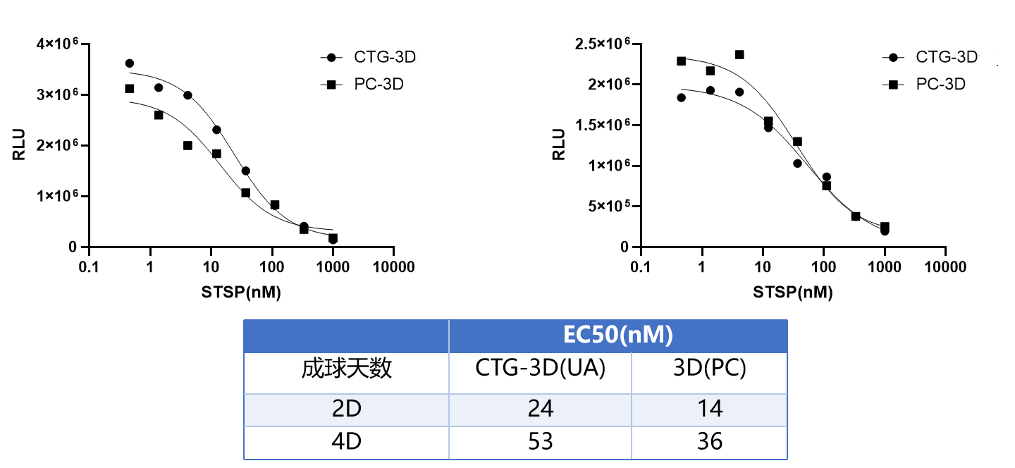
- Experimental Data Analysis: Typical experimental data will show a dose-response relationship. For example, after treating pre-cultured Hela cell 3D spheroids with different concentrations of staurosporine, the measured luminescence signal intensity decreases characteristically with increasing compound concentration, allowing calculation of inhibition rates and half-maximal inhibitory concentration (IC50).
VI. Summary
The UA-Glo® 3D Cell Viability Assay Kit provides a reliable and convenient tool for quantitatively assessing the number of viable cells in 3D culture systems. Its principle of detecting ATP content to reflect cell viability is clear, the operational process is simple and efficient, and the reagent performance is stable with high data quality. The application of this technology facilitates the in-depth development of 3D cell models in drug development, tumor biology, and regenerative medicine, providing strong support for researchers to obtain more biologically relevant experimental data.
VII. Which Manufacturers Provide the UA-Glo® 3D Cell Viability Assay Kit?
Nanjing UA-Bio Technology Co., Ltd. (UA-Bio) has independently developed the "UA-Glo® 3D Cell Viability Assay", a high-performance bioluminescence detection platform specifically designed for viability assessment in three-dimensional cell culture models. This kit is optimized for complex culture systems such as 3D cell spheroids, organoids, and tissue engineering constructs, aiming to accurately and efficiently evaluate the number of viable cells and changes in cell viability in 3D culture systems. It provides stable and reliable standardized solutions for research in tumor microenvironments, drug screening, and regenerative medicine.
| Core Product Advantages |
|---|
| Optimized 3D Tissue Penetration: The kit employs a specially formulated lysis enhancement system that efficiently penetrates the deep structures of 3D cell spheroids, organoids, and scaffold materials, ensuring complete ATP release from the core regions of cell spheroids for precise quantification of 3D culture systems. Its optimized detection reagents and luminescence system maintain excellent detection sensitivity and a wide linear range even in complex matrices. |
| Exceptional Batch-to-Batch Consistency and Stability: Relying on an internationally leading luminescence detection technology platform and standardized production processes, combined with a stringent quality control system, the kit components exhibit high purity, excellent long-term stability, and outstanding batch-to-batch consistency. This provides solid and reliable quality assurance for long-term and continuous 3D culture research. |
| Homogeneous, Wash-Free, Flexible, and Efficient Experimental Mode: The kit adopts a simple "add-lyse-incubate-read" homogeneous operation mode, requiring only the addition of a single detection reagent to the culture wells without cumbersome separation or washing steps. The operational process is extremely simple, greatly reducing experimental time and operational errors. Its optimized formulation system is compatible with multi-well plates (96/384 wells) and high-throughput automated platforms, flexibly suitable for various applications such as 3D tumor spheroid drug screening, organoid toxicity evaluation, and tissue engineering research. |
| Stable and Persistent Luminescence Signal: The kit is specially formulated to produce uniform and stable "glow-type" luminescence signals with a half-life of several hours, providing ample time for large-scale 3D sample detection and automated operations, ensuring the reliability and repeatability of experimental data. |
| Complete Solutions and Professional Support: We provide fully validated standard experimental protocols, typical dose-response curves, and detailed result interpretation guidelines to help you quickly establish stable and reproducible experimental workflows. Nanjing UA-Bio's professional technical team offers comprehensive technical consultation and support for your research design, experimental optimization, and data analysis. |
Nanjing UA-Bio Technology Co., Ltd. is committed to providing cutting-edge, high-quality core reagents and tools for immunology, cell therapy, and innovative drug development. For detailed technical parameters, validation data, or specific application inquiries regarding the "UA-Glo® 3D Cell Viability Assay" (Catalog No.: UA079011), please feel free to contact us.
This article is reviewed and published by the technical expert team of UA
Disclaimer: This article partially utilizes artificial intelligence assistance in its creation. If any content involves copyright or intellectual property issues, please let us know and we promise to verify and remove it as soon as possible.
Purchase recombinant protein, choose Nanjing UA-Bio
UA protein focuses on providing various protein reagents, raw materials, and services required for drug research and development, cell therapy, gene therapy, and basic scientific research, including drug target proteins, immune checkpoint proteins, cytokines, tool enzymes, customized protein expression, and full-length transmembrane protein development. Youai is committed to providing customers with high-quality products and professional services, and building a High-tech Biological Enterprise with International Competitiveness.







 Business license (three certificates in one)