





Lactate Dehydrogenase (LDH) is a stable enzyme widely present in the cytoplasm of all cells. When the cell membrane is intact, LDH remains confined within the cell. However, upon membrane damage or cell death, LDH rapidly leaks into the extracellular culture medium, where it maintains its activity for an extended period. Therefore, by measuring the amount of LDH released into the medium, the extent of cell membrane damage can be precisely quantified, indirectly assessing the cytotoxic effects of external stimuli or compounds.
Traditional LDH assays primarily rely on colorimetric principles: LDH catalyzes the oxidation of lactate to pyruvate, simultaneously reducing NAD⁺ to NADH. NADH, with the aid of lipoamide dehydrogenase, reduces colorless INT to red formazan, which is then measured at 490 nm using a microplate reader.
While this method is classic and reliable, its limitations are evident. First, the sensitivity of colorimetric assays is relatively limited—even with optimized systems, they can only detect LDH released from approximately 500 necrotic cells. For experiments involving precious samples like primary cells or stem cells, or for assessing cytotoxicity in high-throughput drug screening platforms (such as 384- or 1536-well plates), the sensitivity of traditional colorimetric methods falls short. Second, each colorimetric assay requires transferring over 50 µL of supernatant, which can deplete culture volume in multi-timepoint experiments, potentially affecting cell growth conditions. Additionally, the linear range of colorimetric assays is narrow, and improper sample dilution can easily exceed the detection range, adding unnecessary complexity to experiments.
The UA-Glo® LDH Luminescence Cytotoxicity Assay Kit (UA079041) represents a breakthrough by transforming the endpoint signal from visible light absorption in traditional colorimetric assays to bioluminescence, achieving a qualitative leap in sensitivity.
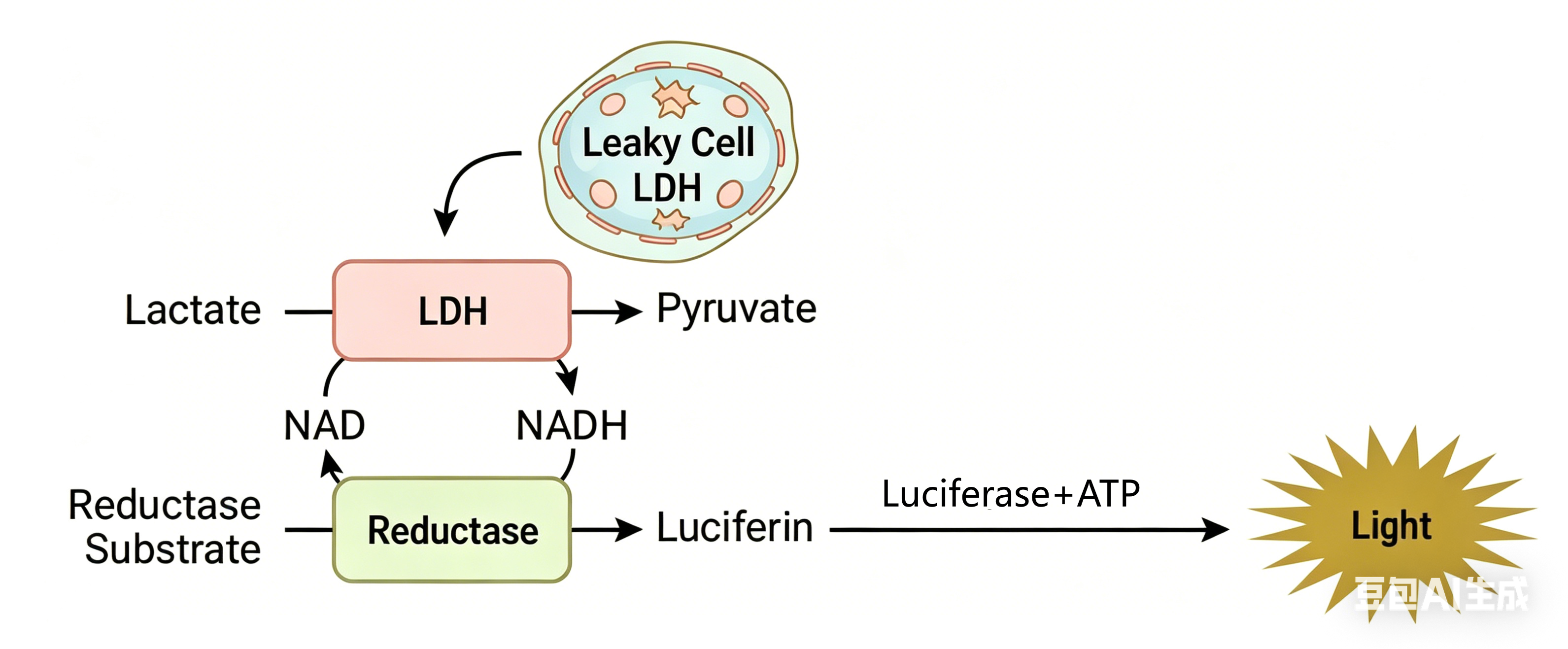 The core principle of the LDH luminescence assay is an elegant enzyme-coupled reaction system. LDH in the sample first catalyzes lactate oxidation while reducing NAD⁺ to NADH. Next, reductase utilizes the generated NADH to convert the reductase substrate into luciferin. Finally, luciferase catalyzes luciferin to emit light. The entire reaction occurs in a single reagent system, with luminescence intensity strictly proportional to LDH activity in the sample.
Compared to traditional colorimetric methods, this "enzyme-coupled luminescence" system offers several key advancements:
- Significantly Improved Sensitivity: Can detect LDH release from fewer than 10 cells—a feat nearly unimaginable with traditional colorimetric methods.
- Minimal Sample Requirement: Only 2-5 µL of cell culture supernatant is needed for detection.
- Homogeneous and Ready-to-Use: UA-Glo® employs a homogeneous, ready-to-use formulation, requiring only a single addition of reagent for detection. This eliminates the multiple manual steps (e.g., supernatant transfer, reaction reagent addition, and incubation) of traditional colorimetric assays, reducing experimental errors.
- No Cell Lysis Required: The assay is non-destructive, allowing cells to continue cultivation post-detection and facilitating compatibility with other cell health assays.
- High Compatibility: Easily scalable to 384- or 1536-well plates, ideal for high-throughput drug screening platforms. Can also be combined with cell viability assays (e.g., CTG kits) or apoptosis detection (e.g., Caspase 3/7) in the same experimental system for multi-dimensional cell health assessment.
- Drug Development: Used for cytotoxicity evaluation (e.g., ADCC) and in vitro activity validation of small-molecule drugs, natural product extracts, PROTAC-targeted protein degraders, antibody-drug conjugates (ADCs), and CAR-T cell therapies.
- 3D Cell Culture: 3D tumor spheroids and organoids mimic in vivo tissue microenvironments more closely, making them more reliable for drug response prediction. However, sample volumes in 3D cultures are often extremely limited, challenging traditional detection methods. The ultra-high sensitivity of luminescent LDH assays enables toxicity studies with time- and dose-dependent drug responses in 3D models using minimal samples.
- Tumor Microenvironment Research: LDH release is an early marker of necrosis, distinct from apoptosis. Combined with apoptosis detection (e.g., Caspase 3/7 assays), researchers can differentiate between drug-induced apoptosis and necrosis, clarifying mechanisms of cell death.
- Nanomaterial and Medical Device Biocompatibility Evaluation: LDH assays are included in industry standards such as YY/T 0993-2015 ("Biological Evaluation of Medical Devices—Nanomaterials: In Vitro Cytotoxicity Tests"), which recommends LDH assays alongside MTT assays.
- Toxicology Assessment: Widely used to determine safe concentration ranges for cosmetics, food additives, and environmental pollutants (e.g., heavy metals, pesticides).
High-performance cell health detection solutions to accelerate drug screening and toxicology research
| Catalog No. | Product Name | Features |
|---|---|---|
| UA079041 | UA-Glo® LDH Luminescence Cytotoxicity Assay | Quantifies extracellular LDH release via coupled reactions involving LDH, reductase, and luciferase to assess cell health. |
| UA070103 | UA-Glo® Luminescent Cell Viability Assay | The "gold standard" for cell viability detection Fastest and most sensitive |
| UA079014 | UA-Glo® Luminescent Cell Viability 2.0 Assay | Upgraded version: single reagent, enhanced stability, ideal for batch processing. |
| UA079015 | UA-Glo® Fluorescent Cell Viability Assay | Fluorescence-based, suitable for multiplex detection. |
| UA079011 | UA-Glo® 3D Cell Viability Assay | Designed for 3D cultures, retaining all features of Luminescent Cell Viability Assay. |
| UA079012 | UA-Glo® Caspase 3/7 Assay | Bioluminescent, filter-free, ultra-sensitive (detects as few as 20 apoptotic cells). |
Note: All UA-Glo products undergo rigorous quality control and are suitable for high-throughput screening and critical mechanistic studies. For technical support, contact UA-Bio.











