





Colorectal cancer is one of the cancers with the highest incidence and mortality rates worldwide, and surgical resection of all tumor tissues with clear margins remains the primary treatment for most colorectal cancer patients. However, complete tumor resection still faces challenges, including incomplete tumor removal, positive resection margins, and peritoneal metastasis. Surgeons primarily rely on visual and tactile diagnosis during surgery, lacking real-time imaging assistance, making it difficult to accurately distinguish adhesions, fibrosis, and tumor lesions. Near-infrared fluorescence imaging is a real-time imaging technology that combines near-infrared fluorescent targeting probes with fluorescence imaging systems to identify specific and highly expressed molecular targets on tumor tissues, differentiating them from normal tissues. Compared to traditional NIR-I, NIR-II significantly overcomes drawbacks such as strong tissue absorption, autofluorescence, and photon scattering, offering high tissue penetration and spatial resolution, which holds promise for precise tumor resection. PE-labeled CEACAM-5/CD66e Fc and Avi-tag proteins can be used to quantitatively detect CEACAM-5 expression levels and binding activity, providing technical tools for colorectal cancer fluorescence imaging research.
The CEACAM-5 gene is overexpressed in approximately 90% of colorectal cancers, with normal tissues expressing CEACAM-5 at levels about 60 times lower than tumor tissues, making it an optimal imaging target for colorectal cancer. In human colorectal cancer cell lines, some cell lines such as HT29-Luc and SW480 overexpress CEACAM-5, while others like colo201 and colo320 do not express CEACAM-5, providing an experimental basis for CEACAM-5 as a targeting marker. Immunofluorescence results show that CEACAM-5 is distributed on the cell membrane surface, and the binding of probes to the cell surface demonstrates the specificity of the targeting effect. Cell binding and blocking experiments further confirm that the targeting probe only binds to cells overexpressing CEACAM-5. These results indicate that CEACAM-5 is an ideal molecular target for targeted imaging of colorectal cancer.
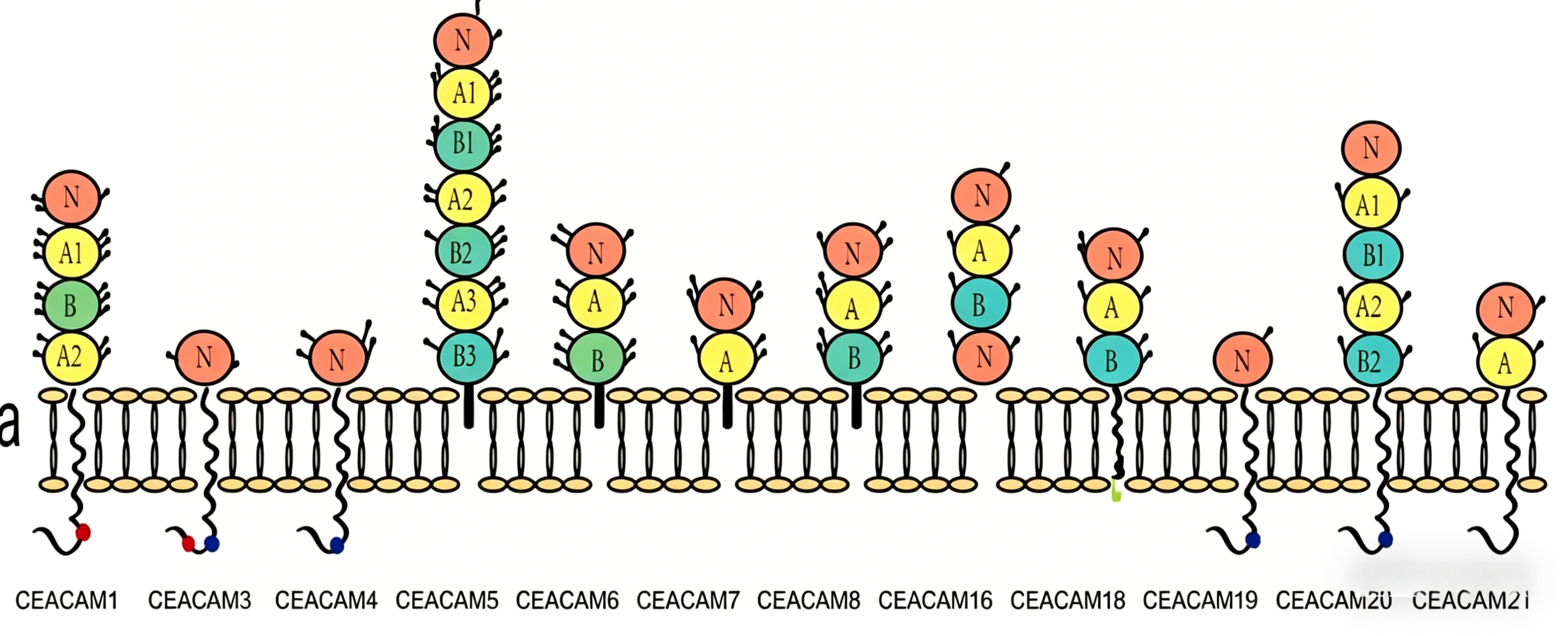
Researchers developed a fluorescent probe constructed by conjugating the near-infrared fluorescent dye IRDye800CW to an anti-CEACAM-5 nanobody. Surface plasmon resonance was used to measure the probe's affinity at different concentrations, with the optimal affinity achieved at a fluorescent molecule-to-protein ratio of 1.25. The probe's absorption spectrum peaked at 776 nm, and its emission spectrum peaked at 789 nm. In the NIR-II range, the probe still exhibited NIR-II signals at 1600 nm, making it a promising NIR-II fluorescent material. The probe's fluorescence intensity in NIR-I or NIR-II showed a linear relationship with concentration, with better performance in NIR-II. PE-labeled CEACAM-5/CD66e Fc and Avi-tag proteins can be used to evaluate the probe's binding ability to CEACAM-5 antigens, validating its targeting specificity.
Researchers compared the performance of 2D5-IRDye800CW in NIR-I and NIR-II imaging of mouse blood vessels and capillaries. Mouse cerebrovascular images at 850 nm (NIR-I) and at 1000 nm and 1200 nm (NIR-II) showed that NIR-II imaging provided clearer visualization of vascular structures compared to NIR-I. Fluorescence cross-sectional intensity distribution of NIR-I/II cerebrovascular images further demonstrated that the probe exhibited stronger fluorescence intensity in NIR-II. Comparisons of mouse hindlimb vessels under different NIR-I/II conditions also confirmed that imaging resolution improved with increasing wavelength. Simulated fluorescence scattering of the probe at a 5 mm depth within fat under NIR-I/II conditions revealed that while both NIR-I and NIR-II exhibited fluorescence scattering, scattering was most severe at 850 nm and almost negligible at 1300 nm. These results indicate that the probe has excellent NIR-II imaging performance.
In in vivo specificity and biodistribution studies, targeting experiments of the probe in HT29-Luc, colo201, and HT29-Luc blocked model mice showed strong fluorescence signals in the kidneys, which weakened over time. The strongest tumor fluorescence intensity was observed 24 hours post-administration, with no fluorescence signal in the control group, confirming the probe's excellent in vivo targeting. The distribution of the probe in various organs 24 hours post-administration revealed that it primarily accumulated in the kidneys, liver, and tumor tissue. BLI-CT 3D reconstruction localized the tumor's position in vivo. Fluorescence signal intensity of intestinal tumors in mice under NIR-I/II conditions showed stronger fluorescence intensity in NIR-II. BLI and HE staining of resected tumors confirmed that the luminescent regions were tumor tissue. In summary, the probe exhibits excellent targeting and, when combined with NIR-II, effectively indicates tumor presence.
In a representative experiment of R0 resection of colorectal cancer guided by NIR-II fluorescence, tumor-bearing mice were administered the probe, and after abdominal exposure, NIR-II fluorescence imaging showed stronger fluorescence signals in the cecum and peritoneum compared to normal tissues, enabling better differentiation between normal and tumor tissues. Under NIR-II fluorescence guidance, in situ colorectal tumor resection was performed, and post-resection NIR-II and BLI imaging confirmed no residual tumor, demonstrating that the probe combined with NIR-II can precisely locate tumor tissue. Compared to NIR-I, NIR-II provided a higher tumor-to-background ratio. In colorectal and peritoneal cancer resection surgeries, NIR-II imaging showed higher fluorescence signals in the cecum and peritoneum than in normal intestinal tissue, correlating with BLI imaging results. Sequential resection of primary tumors, peritoneal metastases, and residual tumors followed by BLI imaging confirmed complete tumor removal. Pathological results and CEACAM-5 immunohistochemical staining of resected tissues confirmed that all removed tissues were tumor tissue.
Nanjing UA-Bio Technology Co., Ltd. (UA-Bio) has independently developed "PE-Labeled CEACAM-5/CD66e Fc&Avi Tag Protein, Human" (Catalog No.: UA011285), a high-performance fluorescently labeled probe specifically designed for carcinoembryonic antigen-related cell adhesion molecule 5 (CEACAM-5) targeting research and solid tumor immunotherapy evaluation. CEACAM-5/CD66e is an important tumor marker for various solid tumors, including colorectal cancer, gastric cancer, and non-small cell lung cancer. This protein consists of the human CEACAM-5 extracellular domain fused with an Fc tag and an Avi tag, labeled with PE fluorescence, enabling efficient binding to anti-CEACAM-5 antibodies or affinity ligands. It serves as a stable and reliable standardized tool for CAR-T cell therapy evaluation, antibody drug screening, tumor marker research, and other fields.
| Core Product Advantages | Detailed Parameters / Functional Description |
|---|---|
| High Purity and Intact Bioactivity | The product employs an internationally leading eukaryotic expression system and highly standardized purification processes, validated through multi-dimensional quality control to ensure >95% purity and correct native conformation (retaining complete glycosylation modifications). The Fc tag enhances protein stability and proper folding, while the Avi tag supports biotinylation applications. The PE labeling process is optimized to ensure high labeling efficiency while maintaining the protein's high-affinity binding to antibodies, accurately simulating the antigenic properties of CEACAM-5 on the surface of solid tumor cells such as colorectal and gastric cancers under physiological conditions.-- |
| High-Brightness PE Labeling | An optimized PE fluorescence labeling ratio ensures stable dye labeling per CEACAM-5 protein molecule, producing bright orange-red fluorescence signals (emission peak ~575 nm) under 488 nm excitation, suitable for flow cytometry (FACS) detection. This provides high sensitivity for evaluating the activity and precise quantification of CEACAM-5-targeting molecules.-- |
| Ideal Tool for Multiple Applications | This protein performs excellently in various applications, including CAR-T cell positivity rate detection by flow cytometry, anti-CEACAM-5 antibody/antagonist screening, competitive binding assays, tumor marker detection and analysis for solid tumors (e.g., colorectal cancer, gastric cancer, non-small cell lung cancer), and flow cytometry. It is widely applicable to CEACAM-5-targeted CAR-T cell therapy development, solid tumor diagnostic tool research, and drug activity evaluation.-- |
| Comprehensive Solutions and Professional Support | We provide thoroughly validated standard experimental protocols, representative flow cytometry data, and detailed result interpretation guidelines to help you quickly establish stable and reproducible CEACAM-5-targeting molecule detection workflows. Nanjing UA-Bio's professional technical team offers full-cycle support for research design, experimental optimization, and data analysis.-- |
Nanjing UA-Bio Technology Co., Ltd. is committed to providing cutting-edge, high-quality core reagents and tools for immunology, cell therapy, and innovative drug development. For detailed technical parameters, validation data, or application consultations regarding "PE-Labeled CEACAM-5/CD66e Fc&Avi Tag Protein, Human" (Catalog No.: UA011285), please feel free to contact us.








