




The core regulatory network of osteoclast differentiation and in vitro induction research tools
Author: Senior Product Manager of UA Views:78 


This article focuses on the central role of osteoclasts in bone metabolic homeostasis, systematically elucidating the synergistic mechanism of the two key cytokines, M-CSF and RANKL, in osteoclast differentiation, and analyzing the regulatory balance of OPG as a natural antagonist.
- Recent Advances
- Product Information
Recent Advances
Core Regulatory Network of Osteoclast Differentiation and In Vitro Induction Research Tools
Overview
This article focuses on the central role of osteoclasts in bone metabolic homeostasis, systematically elaborating the synergistic mechanism of two key cytokines, M-CSF and RANKL, in osteoclast differentiation, and analyzing the regulatory balance of OPG as a natural antagonist.
This article focuses on the central role of osteoclasts in bone metabolic homeostasis, systematically elaborating the synergistic mechanism of two key cytokines, M-CSF and RANKL, in osteoclast differentiation, and analyzing the regulatory balance of OPG as a natural antagonist.
1. The Central Role of Osteoclasts in Bone Metabolic Homeostasis
Human bones are dynamically renewing tissues, with approximately 10% of the bone undergoing remodeling annually in adults. Bone remodeling involves two aspects: bone resorption and bone formation, which are tightly coupled in time and space to maintain normal bone metabolism. Osteoclasts are currently the only known cells in the human body capable of bone resorption. They first form a resorption lacuna on the bone surface, followed by osteoblasts forming new bone within this lacuna, completing the renewal of bone units. When this balance is disrupted—when bone resorption exceeds bone formation—it can lead to common bone metabolic diseases such as osteoporosis and periprosthetic osteolysis. Therefore, studying the differentiation mechanisms of osteoclasts has significant physiological and pathological implications.
However, osteoclasts are scarce in normal human tissues and have a short lifespan, making direct isolation and purification from bone tissues challenging, which has long hindered research progress in this field. It was not until the establishment of in vitro osteoclast-like cell models that researchers were able to systematically analyze the molecular mechanisms of osteoclast differentiation.
2. Two Key Cytokines Regulating Osteoclast Differentiation
Numerous cytokines are involved in osteoclast differentiation, but macrophage colony-stimulating factor (M-CSF) and receptor activator of nuclear factor kappa-B ligand (RANKL) are recognized as the two most critical and essential factors.
1. Macrophage Colony-Stimulating Factor: The "Permissive" Signal for Differentiation
M-CSF can be produced by various cells, including macrophages, endothelial cells, fibroblasts, osteoblasts, and mesenchymal cells. Its biological effects are achieved by binding to the c-fms receptor on the membrane of osteoclast precursors. Upon binding, M-CSF activates the tyrosine kinase activity of c-fms, promoting the survival and proliferation of osteoclast precursors. M-CSF gene-deficient op/op mice exhibit congenital osteopetrosis, with a lack of osteoclasts in vivo. However, after injecting M-CSF into the bone marrow cavity, the number of osteoclasts significantly increases, directly demonstrating the indispensability of M-CSF in osteoclast generation.
M-CSF can be produced by various cells, including macrophages, endothelial cells, fibroblasts, osteoblasts, and mesenchymal cells. Its biological effects are achieved by binding to the c-fms receptor on the membrane of osteoclast precursors. Upon binding, M-CSF activates the tyrosine kinase activity of c-fms, promoting the survival and proliferation of osteoclast precursors. M-CSF gene-deficient op/op mice exhibit congenital osteopetrosis, with a lack of osteoclasts in vivo. However, after injecting M-CSF into the bone marrow cavity, the number of osteoclasts significantly increases, directly demonstrating the indispensability of M-CSF in osteoclast generation.
2. RANKL: The Decisive Signal Driving Terminal Differentiation
RANKL belongs to the tumor necrosis factor superfamily and is primarily expressed by osteoblasts and stromal cells. The promoter region of RANKL contains binding sites for core binding factor-1 (Cbf-1), a key transcription factor for osteoblast differentiation, making Cbf-1 a molecular link between osteoblasts and osteoclasts.
RANKL belongs to the tumor necrosis factor superfamily and is primarily expressed by osteoblasts and stromal cells. The promoter region of RANKL contains binding sites for core binding factor-1 (Cbf-1), a key transcription factor for osteoblast differentiation, making Cbf-1 a molecular link between osteoblasts and osteoclasts.
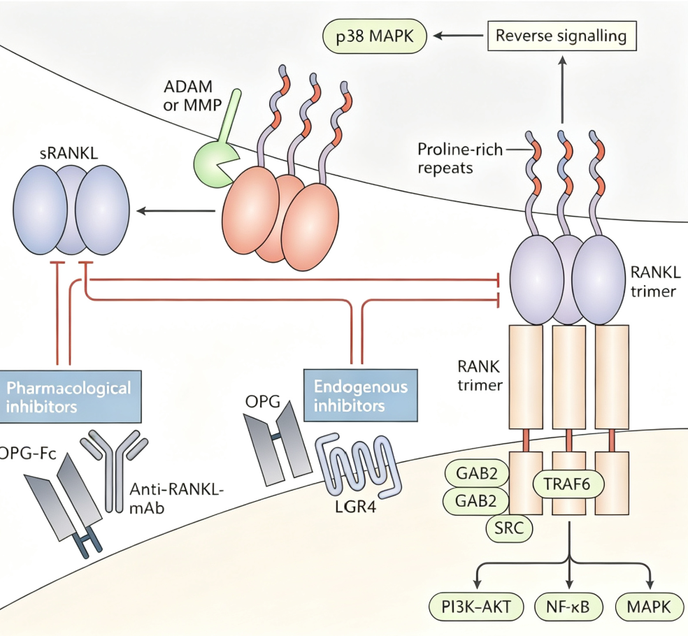
RANKL exists in both membrane-bound and soluble forms. It binds to the RANK receptor on the surface of osteoclast precursors, recruiting TNF receptor-associated factors (especially TRAF6) and activating downstream signaling pathways such as NF-κB and JNK, initiating the transcription of osteoclast-specific genes and driving the fusion of precursor cells into mature multinucleated osteoclasts.
3. Regulatory Balance of the OPG/RANK/RANKL System
Osteoprotegerin (OPG) is also a member of the TNF receptor superfamily and is a secreted glycoprotein primarily synthesized by osteoblasts. OPG can competitively bind to RANKL with a higher affinity than RANK, effectively blocking the RANK/RANKL signaling pathway and inhibiting excessive osteoclast differentiation. The RANKL/OPG ratio determines the strength of osteoclast differentiation—an increased ratio promotes osteoclast generation, while a decreased ratio inhibits differentiation. Under pathological conditions such as estrogen deficiency, glucocorticoid excess, or inflammatory cytokine stimulation, the RANKL/OPG ratio increases, leading to excessive osteoclast activation, which is a core mechanism of postmenopausal osteoporosis and bone erosion in rheumatoid arthritis.
4. Applications of In Vitro Osteoclast Differentiation Models
Over the past decade, the establishment of in vitro osteoclast induction differentiation models has significantly advanced research in this field. The basic principle is that under the synergistic action of M-CSF and RANKL, human mononuclear cells (such as the THP-1 cell line or peripheral blood mononuclear cells) can be directionally differentiated into multinucleated osteoclasts with bone resorption function in vitro. In experimental operations, M-CSF supports the survival and proliferation of precursor cells, while RANKL drives terminal differentiation and fusion, with both being indispensable.
5. Conclusion
Osteoclasts are the sole executors of bone resorption, and their differentiation is tightly controlled by a precise regulatory network centered on M-CSF and RANKL, with OPG as a natural antagonist. Dysregulation of this signaling axis is a common pathological basis for various bone metabolic diseases. The in vitro osteoclast induction differentiation model, utilizing M-CSF and RANKL as key cytokines, provides an important experimental platform for in-depth research on osteoclast biology and screening of anti-resorptive drugs. Uni offers a human monocyte osteoclast differentiation cytokine kit, which includes recombinant human M-CSF and recombinant human RANKL—both highly active, high-purity, and low-endotoxin cytokines—that efficiently drive human precursor cells to differentiate into mature osteoclasts. This kit is suitable for bone metabolism mechanism research, drug screening, and disease model construction related to osteoporosis.
This article is reviewed and published by the technical expert team of UA
Disclaimer: This article partially utilizes artificial intelligence assistance in its creation. If any content involves copyright or intellectual property issues, please let us know and we promise to verify and remove it as soon as possible.
Purchase recombinant protein, choose Nanjing UA-Bio
UA protein focuses on providing various protein reagents, raw materials, and services required for drug research and development, cell therapy, gene therapy, and basic scientific research, including drug target proteins, immune checkpoint proteins, cytokines, tool enzymes, customized protein expression, and full-length transmembrane protein development. Youai is committed to providing customers with high-quality products and professional services, and building a High-tech Biological Enterprise with International Competitiveness.
Target proteins | membrane proteins | cytokines | enzymes | viral antigens | protein customization
Nanjing UA Biotechnology Co., Ltd. Email:order@ua-bio.com Phone:+86-25-56221161

Product Information

Link:





©2021-2026 all rights reserved UA Biotechnology Co.,Ltd.
Privacy Policy 
网站备案号 浙ICP备2022019033号-2 浙公网安备33010202004867号
 Business license (three certificates in one)
 Tel:+86-25-56221161
E-mail:order@ua-bio.com
All products sold on this website shall not be used for clinical diagnosis or treatment of humans or animals, and shall only be used for non-medical purposes such as industrial or scientific research.
