




Caspase cascade reaction: From initiation to execution of the apoptotic signaling network and high-throughput detection applications
Author: Senior Product Manager of UA Views:133 


This article focuses on the central role of the Caspase family of proteases in apoptotic signal transduction, systematically elucidating the cascade reaction mechanisms through which they are activated via the intrinsic and extrinsic pathways. It analyzes the hierarchical functional分工 between initiator Caspases and effector Caspases, as well as the cleavage effects on key downstream substrates.
- Recent Advances
- Product Information
Recent Advances
Caspase Cascade Reaction: From Initiation to Execution of the Apoptotic Signaling Network and High-Throughput Detection Applications
Overview
This article focuses on the central role of the Caspase family of proteases in apoptotic signal transduction, systematically elucidating the cascade activation mechanisms through both intrinsic and extrinsic pathways, and analyzing the hierarchical functional分工 between initiator Caspases and effector Caspases as well as the cleavage effects on downstream key substrates.
This article focuses on the central role of the Caspase family of proteases in apoptotic signal transduction, systematically elucidating the cascade activation mechanisms through both intrinsic and extrinsic pathways, and analyzing the hierarchical functional分工 between initiator Caspases and effector Caspases as well as the cleavage effects on downstream key substrates.
I. The Caspase Family: Core Executors of Apoptotic Signal Transduction
Caspases are a highly conserved family of cysteine proteases, named for their catalytic activity dependent on cysteine residues and their specific cleavage ability after aspartic acid residues. In the cascade reaction of apoptotic signal transduction, different Caspase members协同作用, forming a precisely regulated protease cleavage network. Once initiated, this cascade leads to the cleavage of numerous critical intracellular proteins, resulting in cell解体 and death, ultimately清除 cell debris through phagocytosis to complete the apoptotic program.
Based on functional and structural characteristics, the Caspase family can be divided into two subgroups: initiator Caspases and effector Caspases. Initiator Caspases include Caspase-8, Caspase-9, Caspase-10, and Caspase-12, which possess long N-terminal prodomains and are activated through specific protein-protein interaction domains (such as death effector domains or CARD domains) that bind to upstream signaling complexes. Effector Caspases include Caspase-3, Caspase-6, and Caspase-7, which exist as inactive zymogens in the cytoplasm and are activated by proteolytic cleavage by initiator Caspases, subsequently cleaving various key substrates to execute the final cell解体 effects.
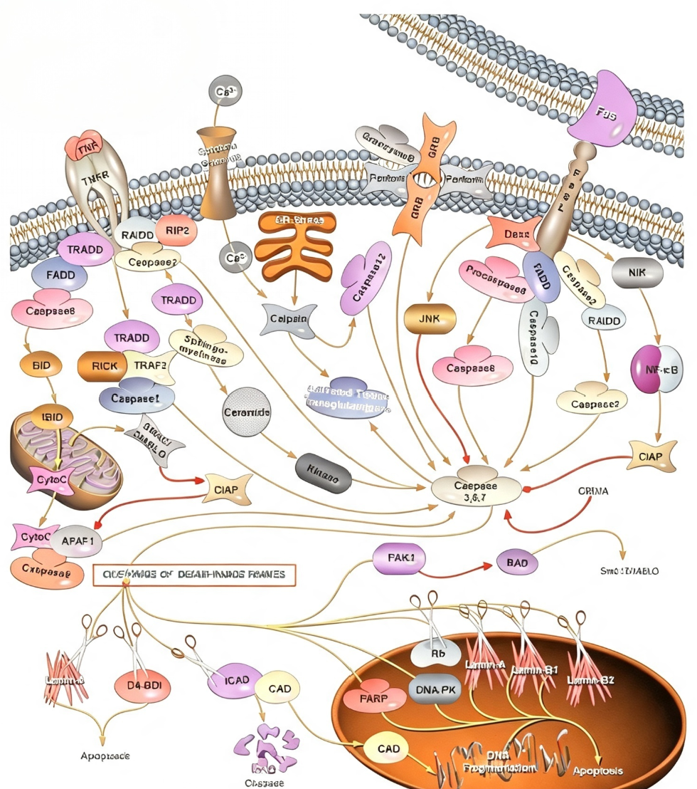
II. Extrinsic Apoptotic Pathway: Signal Initiation Mechanism via Cell Surface Receptors
The extrinsic apoptotic pathway is mediated by cell surface death receptors, primarily involving the activation of Caspase-8 and Caspase-10. Binding of Fas ligand to the Fas receptor recruits pro-Caspase-8 via the adaptor protein FADD, forming the death-inducing signaling complex (DISC), which leads to auto-cleavage and activation of Caspase-8. Similarly, binding of tumor necrosis factor (TNF) to the TNFR1 receptor initially forms a complex containing adaptor proteins such as TRADD, RIP, and TRAF2, which then recruits FADD and pro-Caspase-8. Activated Caspase-8 directly cleaves and activates downstream effector Caspases-3 and -7, while also cleaving BID to generate truncated tBID, transmitting the apoptotic signal to the mitochondrial pathway.
Activation of the extrinsic pathway not only induces apoptosis but also involves complex cross-regulation with survival signals. TNF signaling can simultaneously activate the NF-κB transcription factor, inducing the expression of anti-apoptotic proteins such as the IAP family, thereby balancing pro-apoptotic and pro-survival signals.
III. Intrinsic Apoptotic Pathway: Mitochondrial Damage and Caspase-9 Activation
The intrinsic apoptotic pathway is triggered by stimuli such as DNA damage, growth factor deprivation, endoplasmic reticulum (ER) stress, and elevated intracellular Ca²⁺ levels. Mitochondria play a central role in this pathway. Under apoptotic stimuli, mitochondrial outer membrane permeability increases, releasing pro-apoptotic factors such as cytochrome c and SMAC/DIABLO. Cytochrome c binds to APAF1 to form the apoptosome, which recruits and activates pro-Caspase-9. Activated Caspase-9 then cleaves pro-Caspase-3, initiating the effector Caspase cascade. SMAC/DIABLO amplifies the apoptotic signal by binding and inhibiting IAP family proteins, thereby解除 IAP-mediated suppression of Caspase activity. Additionally, ER stress can activate Caspase-12 via Ca²⁺ signaling, representing an independent initiation pathway.
IV. Downstream Substrate Cleavage by Effector Caspases-3/7 and Apoptotic Execution
Caspase-3 and Caspase-7 are the most critical effector molecules in the apoptotic cascade, both recognizing and cleaving the same DEVD tetrapeptide sequence. Once activated by initiator Caspases, they execute the apoptotic program by cleaving a series of key substrates. At the cytoskeletal level, Caspase-3/7 cleave structural proteins such as fodrin and lamin, leading to cytoskeletal解体 and nuclear membrane rupture. In DNA metabolism, Caspase-3 cleaves ICAD, releasing CAD nuclease to mediate DNA fragmentation—a hallmark biochemical feature of apoptotic cells. Simultaneously, Caspase-3/7 also cleave regulatory proteins such as PARP (involved in DNA damage repair), DNA-PK, and Rb,彻底阻断 cellular repair and proliferation capabilities.
V. Caspase-3/7 Activity Detection: Key Technologies from Basic Mechanisms to Drug Development
In apoptosis mechanism research, anticancer drug screening, and cytotoxicity assessment, sensitive and reliable Caspase-3/7 activity detection methods are indispensable core tools. Traditional methods such as Western blot, immunohistochemistry, or fluorescence-based assays often have limitations in sensitivity, throughput, or operational convenience. To address this need, UA offers the UA-Glo® Caspase 3/7 Assay. This kit employs a homogeneous chemiluminescent detection system, with its core design featuring a highly specific luminescent substrate for Caspase-3/7—containing a DEVD sequence linked to an aminoluciferin precursor molecule. Activated Caspase-3/7 specifically cleaves the DEVD sequence, releasing aminoluciferin, which generates a "glow-type" chemiluminescent signal catalyzed by an optimized luciferase system. The signal intensity is proportional to Caspase-3/7 activity. The kit features a "add-mix-detect" homogeneous操作模式, requiring no washing or cell separation steps, and is compatible with 96/384-well plate high-throughput formats. The luminescent signal is stable with a half-life exceeding 2 hours, and the Z′ factor typically exceeds 0.7.
VI. Conclusion
The Caspase cascade reaction, through the精密协同 of both extrinsic and intrinsic activation pathways, amplifies apoptotic signals stepwise, ultimately executing关键 substrate cleavage via effector Caspases-3/7 to complete cell解体. Dysregulation of this signaling network is closely linked to various major diseases, and深入理解 its activation and regulatory mechanisms holds significant fundamental and clinical translational importance. High-sensitivity, high-throughput-compatible Caspase-3/7 activity detection technologies will continue to play an indispensable role in apoptosis mechanism解析, drug discovery, and safety evaluation.
This article is reviewed and published by the technical expert team of UA
Disclaimer: This article partially utilizes artificial intelligence assistance in its creation. If any content involves copyright or intellectual property issues, please let us know and we promise to verify and remove it as soon as possible.
Purchase recombinant protein, choose 
UA protein focuses on providing various protein reagents, raw materials, and services required for drug research and development, cell therapy, gene therapy, and basic scientific research, including drug target proteins, immune checkpoint proteins, cytokines, tool enzymes, customized protein expression, and full-length transmembrane protein development. Nanjing Univ is committed to providing customers with high-quality products and professional services, and building a High-tech Biological Enterprise with International Competitiveness.
Target proteins | membrane proteins | cytokines | enzymes | viral antigens | protein customization
Nanjing UNIV Biotechnology Co., Ltd. Email:order@ua-bio.com Phone:+86-25-56221161

Product Information

Link:





©2021-2026 all rights reserved UNIV Biotechnology Co.,Ltd.
Privacy Policy
网站备案号 苏ICP备2025207586号-2 浙公网安备33010202004867号
 Business license (three certificates in one)
 Tel:+86-25-56221161
E-mail:order@ua-bio.com
All products sold on this website shall not be used for clinical diagnosis or treatment of humans or animals, and shall only be used for non-medical purposes such as industrial or scientific research.


