




H-2Db MHC Tetramer: The Innovation and Application Expansion of Antigen - specific T Cell Detection Technology


Antigen - specific T cells are central effector cells of the adaptive immune system. They play a key role in viral infection, tumor immunosurveillance, and vaccine - induced protective immunity. Traditional detection methods, limited by low sensitivity or complex procedures, struggle to precisely quantify low - frequency antigen - specific T cells. The H-2Db MHC tetramer technology has broken through this barrier. By forming high - affinity polymeric complexes, it enables single - cell detection and functional analysis of CD8+ T cells.
- Recent Advances
- Product Information
H-2Db MHC Tetramer
Introduction
Antigen-specific T cells, as core effector cells of the adaptive immune system, play pivotal roles in viral infections, tumor immunosurveillance, and vaccine-induced protective immunity. Traditional detection methods, limited by insufficient sensitivity or operational complexity, struggle to accurately quantify low-frequency antigen-specific T cells. The emergence of H-2Db MHC tetramer technology has overcome this bottleneck by enabling single-cell resolution detection and functional analysis of CD8+ T cells through high-affinity multimeric complexes. This article systematically elucidates the preparation workflow, technical principles of H-2Db tetramers, and their applications in the lymphocytic choriomeningitis virus (LCMV) infection model, while exploring the multidimensional value of this technology in immunological research.
H-2Db Tetramer Preparation Technology Platform
1. Genetic Engineering of Core Components
H-2Db heavy chain cDNA was cloned via RT-PCR to construct a fusion protein expression vector containing the H-2Db extracellular domain and a biotin ligase BirA substrate peptide (BSP). This design achieves two critical functions:
- Conformational Integrity: Retention of the H-2Db α1/α2 domain peptide-binding groove ensures specific interaction with target epitopes.
- Enzymatic Tagging Site: The C-terminal BSP sequence enables subsequent biotinylation for tetramer formation.
2. Prokaryotic Expression System Optimization
The recombinant vector was introduced into E. coli BL21(DE3) for high-yield soluble protein production. Optimized induction conditions included:
- Strain: BL21(DE3)
- Induction: 0.5 mM IPTG, 25°C shaking culture for 16 hours
- Yield: ~15 mg fusion protein per liter of culture
3. In Vitro Refolding and Peptide Loading
Correct assembly of H-2Db heavy chain with human β2-microglobulin (β2m) was facilitated by dilution refolding in the presence of LCMV GP33-41 epitope peptide (KAVYNFATM, KAV). Key steps included:
- Refolding Buffer: 50 mM Tris-HCl, 100 mM NaCl, 2 mM EDTA, 5 mM GSH, 0.5 mM GSSG, pH 8.0
- Gradient Dialysis: Sequential dialysis against 4 M, 2 M, and 0 M urea buffers (48 hours total)
- Purification: Size-exclusion chromatography (Superdex 200) yielded >95% pure H-2Db/KAV monomers
4. Tetramer Assembly and Labeling
Biotinylated monomers were tetramerized by mixing with streptavidin-PE (SA-PE) at a 4:1 molar ratio, followed by 1-hour incubation at room temperature. Final purification via gel filtration removed free monomers, yielding uniform H-2Db/KAV-PE tetramers.
Table 1. Key Parameters for H-2Db Tetramer Preparation
| Step | Key Reagents/Conditions | Output Metrics |
|---|---|---|
| Gene Cloning | H-2Db cDNA, BSP sequence | Recombinant expression vector |
| Protein Expression | BL21(DE3), 0.5 mM IPTG | 15 mg/L soluble protein |
| In Vitro Refolding | GP33-41 peptide, β2m | >85% correctly folded monomers |
| Biotinylation | BirA enzyme, 50 μM biotin | >90% labeling efficiency |
| Tetramer Assembly | SA-PE (4:1 ratio) | Fluorescently labeled tetramer |
Application of H-2Db Tetramers in LCMV-Specific CD8+ T Cell Detection
1. Animal Model and Immunization Protocol
C57BL/6 mice (H-2Db phenotype) were subcutaneously injected with 1×10⁵ PFU LCMV Armstrong strain to establish acute infection. Peripheral blood, spleen, and draining lymph nodes were harvested at day 8 post-infection (peak immune response).
2. Flow Cytometry Detection Workflow
- Sample Processing: Red blood cell lysis, cell resuspension in 2% FBS/PBS at 1×10⁷/mL
- Tetramer Staining: 1 μg H-2Db/KAV-PE tetramer per tube, 4°C incubation for 1 hour
- Co-Staining: Anti-CD8-APC, anti-CD44-FITC antibodies (30-minute incubation)
- Analysis: CD8+ gating, with tetramer+CD44+ cells defined as antigen-specific
3. Results Interpretation
- Tissue Distribution:
- Peripheral blood: 0.27% KAV-specific CD8+ T cells
- Spleen: 0.24%
- Draining lymph nodes: 0.11%
- Phenotypic Analysis: >95% tetramer+ cells expressed activation marker CD44 (effector memory phenotype)
- Functional Validation: Intracellular cytokine staining (ICS) confirmed IFN-γ and TNF-α production in 85% of tetramer+ cells
Table 2. Frequency of LCMV-Specific CD8+ T Cells in Different Tissues
| Tissue | Detection Frequency (%) | Fluorescence Intensity (MFI) | Cell Viability (%) |
|---|---|---|---|
| Peripheral Blood | 0.27 ± 0.03 | 12,450 ± 870 | 92.1 ± 1.4 |
| Spleen | 0.24 ± 0.02 | 9,820 ± 650 | 85.7 ± 2.1 |
| Draining LN | 0.11 ± 0.01 | 7,310 ± 520 | 78.3 ± 3.0 |
Technical Advantages and Clinical Translation Potential
1. Methodological Innovations
- Sensitivity Enhancement: Detects antigen-specific T cells at frequencies as low as 0.001% (vs. ELISPOT limit of ~0.01%)
- Phenotypic-Functional Integration: Enables simultaneous analysis of differentiation status (e.g., CD62L, CD127) and effector functions (perforin, granzyme B) via multicolor flow cytometry
2. Disease Model Expansion
- Oncoimmunology: Tracked PD-1 inhibitor-induced tumor-infiltrating lymphocyte (TIL) expansion in MC38 colon cancer models using H-2Db/AH1-A5 tetramers
- Autoimmunity: Revealed pathogenicity of myelin oligodendrocyte glycoprotein (MOG)-specific T cells in EAE models via H-2Db/MOG35-55 tetramers
3. Technological Iterations
- Flexible Peptide Exchange Platform: UV-mediated peptide exchange enables rapid screening of multiple epitopes using a single tetramer batch
- Multi-Omics Integration: Coupling with CITE-seq allows single-cell TCR sequencing, phenotypic profiling, and functional assessment
Conclusion
The H-2Db MHC tetramer technology provides a transformative tool for antigen-specific T cell research through precise molecular engineering and optimized detection strategies. Its successful application in LCMV infection models has deepened understanding of virus-specific immune dynamics while pioneering new avenues for vaccine evaluation, tumor immunotherapy monitoring, and autoimmune pathogenesis studies. As multidisciplinary technologies converge, H-2Db tetramers will continue to advance precision immunology, heralding the era of personalized medicine.
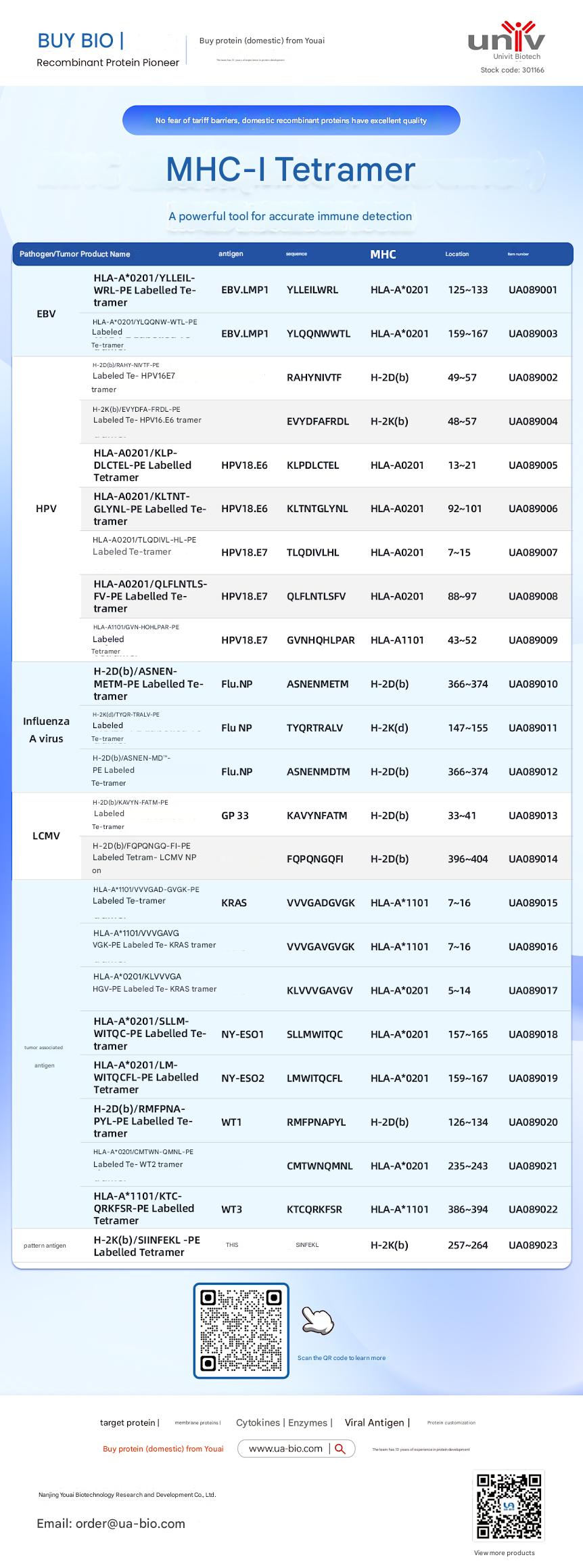
| Disease Category | Product Name | Antigen | Sequence | MHC | Position | Product Number |
|---|---|---|---|---|---|---|
| EBV | HLA-A*0201/YLELLVWRL-PE Labelled Tetramer | EBV.LMP1 | YLELLVWRL | HLA-A*0201 | 125-133 | UA089001 |
| EBV | HLA-A*0201/YLQQNWTL-PE Labelled Tetramer | EBV.LMP1 | YLQQNWTL | HLA-A*0201 | 159-167 | UA089003 |
| EBV | H-2Db(b)/RAHY-NIVTF-PE Labelled Tetramer | HPV16.E7 | RAHYNIVTF | H-2Db | 49-57 | UA089002 |
| HPV | H-2K(b)/EVYDFA-FRQL-PE Labelled Tetramer | HPV16.E6 | EVYDFARDL | H-2Kb | 48-57 | UA089004 |
| HPV | HLA-A*0201/KLP-DLCTL-PE Labelled Tetramer | HPV18.E6 | KLPDCTL | HLA-A*0201 | 13-21 | UA089005 |
| HPV | HLA-A*0201/KLTNT-GLYQL-PE Labelled Tetramer | HPV18.E6 | KLTNTGLYNL | HLA-A*0201 | 92-101 | UA089006 |
| HPV | HLA-A*0201/TLODIVIHL-PE Labelled Tetramer | HPV18.E7 | TLODIVIHL | HLA-A*0201 | 7~15 | UA089007 |
| HPV | HLA-A*0201/QFLNTL-FV-PE Labelled Tetramer | HPV18.E7 | QFLNTLFSV | HLA-A*0201 | 88-97 | UA089008 |
| HPV | HLA-A*1101/GVNHQLPAR-PE Labelled Tetramer | HPV18.E7 | GVNHQLPAR | HLA-A*1101 | 43-52 | UA089009 |
| Influenza A Virus | H-2D(b)/ASNENMETM-PE Labelled Tetramer | Flu.NP | ASNENMETM | H-2Db | 366-374 | UA089010 |
| Influenza A Virus | H-2K(d)/TYQR-TRALY-PE Labelled Tetramer | Flu.NP | TYQRTRALY | H-2Kd | 147-155 | UA089011 |
| Influenza A Virus | H-2D(b)/ASNEN-MDTM-PE Labelled Tetramer | Flu.NP | ASNENMDTM | H-2Db | 366-374 | UA089012 |
| LCMV | H-2D(b)/KAVYNFATM-PE Labelled Tetramer | GP 33 | KAVYNFATM | H-2Db | 33-41 | UA089013 |
| LCMV | H-2D(b)/FQPGQGFVK-PE Labelled Tetramer | LCMV NP | FQPGQGFVK | H-2Db | 396-404 | UA089014 |
| Tumor-related | HLA-A*1101/VVGADGVK-PE Labelled Tetramer | KRAS | VVGADGVK | HLA-A*1101 | 7~16 | UA089015 |
| Tumor-related | HLA-A*1101/VVGAGVGK-PE Labelled Tetramer | KRAS | VVGAGVGK | HLA-A*1101 | 7~16 | UA089016 |
| Tumor-related | HLA-A*0201/KLVVGAGV-PE Labelled Tetramer | KRAS | KLVVGAGV | HLA-A*0201 | 5~14 | UA089017 |
| Tumor-related | HLA-A*0201/SLLMWITQC-PE Labelled Tetramer | NY-ESO1 | SLLMWITQC | HLA-A*0201 | 157-165 | UA089018 |
| Melanoma | HLA-A*0201/LMWITQCFL-PE Labelled Tetramer | NY-ESO2 | LMWITQCFL | HLA-A*0201 | 159-167 | UA089019 |
| Melanoma | H-2Db(b)/MMFPNA-P1-PE Labelled Tetramer | WT1 | RMFPNAPL | H-2Db | 126-134 | UA089020 |
| Melanoma | HLA-A*0201/CMTWV-PE Labelled Tetramer | WT2 | CMTWVNMDM | HLA-A*0201 | 235-243 | UA089021 |
| Melanoma | HLA-A*1101/KTCQRKSF-PE Labelled Tetramer | WT3 | KTCQRKSF | HLA-A*1101 | 386-394 | UA089022 |
| Ovarian Cancer | H-2K(b)/SINFEKL-PE Labelled Tetramer | OVA | SINFEKL | H-2Kb | 257-264 | UA089023 |
This article is reviewed and published by the technical expert team of UA
Disclaimer: This article partially utilizes artificial intelligence assistance in its creation. If any content involves copyright or intellectual property issues, please let us know and we promise to verify and remove it as soon as possible.
Purchase recombinant protein, choose Nanjing UA-Bio
UA protein focuses on providing various protein reagents, raw materials, and services required for drug research and development, cell therapy, gene therapy, and basic scientific research, including drug target proteins, immune checkpoint proteins, cytokines, tool enzymes, customized protein expression, and full-length transmembrane protein development. Youai is committed to providing customers with high-quality products and professional services, and building a High-tech Biological Enterprise with International Competitiveness.







 Business license (three certificates in one)