Melanoma MHC Tetramer: Decoding Mechanisms of Tumor Immune Evasion and Novel Strategies for Precision Therapy
Immunotherapy has emerged as one of the cornerstone paradigms in cancer treatment, with innovative therapies such as immune checkpoint inhibitors (ICIs) significantly improving survival outcomes for patients with metastatic cancer. Despite the breakthrough progress made by immunotherapy, particularly represented by ICIs, clinical practice reveals that most patients still confront challenges of primary/adaptive resistance or acquired resistance, alongside persistently high incidence rates of treatment-related immune-related adverse events (irAEs).
- Recent Advances
- Product Information
Recent Advances
Melanoma MHC Tetramer
Abstract
Immunotherapy has become a core paradigm in cancer treatment, with innovative therapies like immune checkpoint inhibitors (ICIs) significantly improving the survival of patients with metastatic cancer. However, most patients still face challenges such as primary/adaptive resistance, acquired resistance, and high rates of immune-related adverse events (irAEs). This highlights the urgent need to understand the molecular mechanisms of anti-tumor immune responses, particularly the precise mechanisms of tumor cell recognition by T cells, which is crucial for identifying biomarkers of therapeutic efficacy/resistance and developing strategies to overcome resistance.
The immune system primarily senses tumor cell heterogeneity through the major histocompatibility complex (MHC) system. While MHC-I molecules are widely expressed in most tumor cells, increasing evidence shows that tumor subpopulations from different tissues can present antigens via MHC-II molecules, thereby regulating the intensity and quality of anti-tumor immune responses. The functional repositioning of MHC-II molecules in anti-tumor immunity is gaining significant academic attention.
Numerous preclinical and clinical studies have confirmed a significant association between tumor-specific MHC-II expression and tumor rejection reactions in cancer patients (including those undergoing immunotherapy) and animal models. Previous research has revealed a positive correlation between tumor cell MHC-II expression levels and the efficacy of anti-PD-1 therapy. This has prompted an in-depth exploration of the biological functions of MHC-II molecules in the tumor microenvironment and their regulatory mechanisms on immunotherapy efficacy. This article focuses on the following core questions: How does MHC-II expression on tumor cells reshape the anti-tumor immune response? What are the key nodes in its regulatory network? Can interventions targeting the MHC-II pathway break through existing immunotherapy bottlenecks? Systematically addressing these scientific questions may provide a theoretical basis for developing next-generation cancer immunotherapies.
MHC-II and MHC-I: Differential Regulation in Cancer Immunotherapy
The clinical breakthroughs of immunotherapies like ICIs have significantly extended the survival of cancer patients, but their efficacy is limited by primary/adaptive resistance and irAEs. Understanding the molecular basis of anti-tumor immune responses, especially T cell recognition of tumor antigens, is strategically important for discovering efficacy prediction biomarkers and overcoming resistance.
Current research reveals that the immune system primarily senses tumor cell heterogeneity through the MHC system. Despite the widespread expression of MHC-I molecules on most nucleated cells, increasing evidence表明 that specific tumor subpopulations can present antigens via MHC-II molecules, thereby regulating anti-tumor immune responses. The central role of MHC-II molecules in CD4⁺ T lymphocyte activation is redefining their value in immunotherapy.
Antigen Presentation Pathways and T Cell Subset Activation Differences
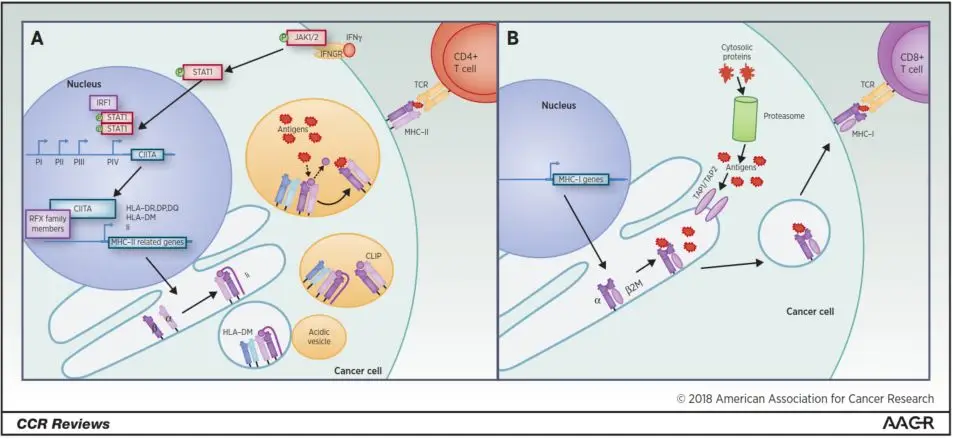
MHC-I Pathway: As an endogenous antigen presentation system, MHC-I molecules present degradation products of cytoplasmically synthesized proteins to CD8⁺ cytotoxic T cells, directly triggering target cell killing. This pathway is the classic route for ICIs, with CD8⁺ T cells being the main effector cells of anti-tumor immunity. MHC-II Pathway: As an exogenous antigen presentation system, MHC-II molecules, primarily expressed by professional antigen-presenting cells (pAPCs), present extracellular protein antigen degradation fragments to CD4⁺ T helper cells. CD4⁺ T cells, through secreting cytokines and providing co-stimulatory signals, have become an indispensable regulatory hub for ICI efficacy.
Structural-Functional Differences Determine Immunomodulatory Role Divergence
MHC-I and MHC-II molecules differ essentially in subunit composition, antigen-binding groove structure, and TCR interaction modes:
Molecular Architecture: MHC-I forms a heterodimer of heavy chain ( - chain) and -microglobulin, with a closed antigen-binding groove accommodating 8 - 10 amino acid residues; MHC-II, composed of - and -chains, has an open-binding groove accommodating 13 - 25 residues. Antigen Spectrum: MHC-I mainly presents endogenous proteins, while MHC-II preferentially displays extracellular proteins. Immune Regulatory Network: CD4⁺ T cells, by recognizing MHC-II-antigenic peptide complexes, not only directly kill MHC-II⁺ tumor cells but also activate pAPCs, upregulate co-stimulatory molecules, and promote CD8⁺ T cell proliferation, constructing multi-layered anti-tumor immune responses.
Clinical research further confirms a positive correlation between tumor cell MHC-II expression levels and anti-PD-1 therapy efficacy, suggesting the MHC-II pathway may be a new target for overcoming immunotherapy resistance.

MHC-II molecules, formed by non-covalent binding of - and -chains, have an open antigen-binding groove accommodating peptide segments of 13 - 25 amino acid residues. This structural characteristic allows them to bind a broader range of proteolytic products than MHC-I, significantly expanding the antigen spectrum recognizable by T cells. MHC-II expression is mainly driven by the class II trans-activator (CIITA), which recruits the RNA polymerase II complex and histone modification enzymes to the MHC-II gene locus for epigenetic regulation.
In contrast, MHC-I molecules are composed of polymorphic -chains and non-polymorphic β₂-microglobulin (β2M) linked by covalent bonds. Their antigen processing and presentation strictly depend on the proteasome-TAP transporter-endoplasmic reticulum axis. Although constitutively expressed in normal tissues, tumor cells can downregulate MHC-I expression via mechanisms like β2M gene deletion or JAK-STAT pathway mutations, evading CD8⁺ T cell recognition. Clinically, MHC-I-deficient tumors often exhibit primary resistance to anti-PD-1 therapy, yet some immune escape variants maintain MHC-II expression, whose functional relevance remains unclear.
Current research suggests that MHC-I and MHC-II are independently regulated in cancer and differentially impact immunotherapy: MHC-I absence directly weakens tumor immune surveillance, while MHC-II, beyond traditional antigen presentation, may activate CD4⁺ T cell-mediated regulatory networks, forming an alternative immune response pathway in specific cancers.
Tumor-specific MHC-II (tsMHC-II): Impact on Cancer Immunotherapy
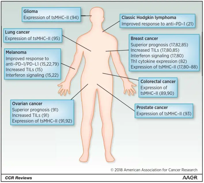
While professional antigen-presenting cells (pAPCs) are the classic site of MHC-II expression, tumor cells can present antigens by aberrantly activating MHC-II pathways. This tumor-specific MHC-II (tsMHC-II) expression has been confirmed in various solid and hematologic malignancies. Its expression level significantly correlates with anti-tumor immune response intensity and clinical prognosis.

Clinical Evidence
Melanoma Treatment Response Prediction Johnson et al. (2016) found bimodal MHC-II expression in 60 melanoma cell lines. Gene set enrichment analysis showed MHC-II⁺ cell lines were enriched for immune-related features. In two independent anti-PD-1 treatment cohorts, tumor cell MHC-II expression correlated positively with objective response rate (ORR), progression-free survival (PFS), and overall survival (OS), and with CD4⁺/CD8⁺ tumor-infiltrating lymphocyte (TIL) density.
Triple-negative Breast Cancer Prognosis Improvement Forero et al. (2016) reported that in triple-negative breast cancer (TNBC) patients, MHC-II antigen presentation pathway expression was an independent prognostic factor. High MHC-II expression was associated with better prognosis, as it activated CD4⁺ T cell-mediated immune surveillance, reducing postoperative recurrence risk.
Classic Hodgkin Lymphoma Treatment Breakthrough Roemer et al. (2018) found in the CheckMate 205 trial that Nivolumab induced a 66% ORR in relapsed/refractory classic Hodgkin lymphoma, with efficacy positively related to MHC-II expression on HRS cells, independent of MHC-I expression.
Pan-cancer Immune Response Regulation Rodig et al. (2018) analyzed 181 treatment-naive melanoma samples:
-
43% had MHC-I loss, predicting primary resistance to anti-CTLA-4 therapy.
-
30% had MHC-II expression (>1% tumor cells), linked to activated IFN-γ signaling and higher anti-PD-1 response (OR = 3.2).
Biological Mechanisms and Clinical Implications
tsMHC-II has multi-dimensional immunomodulatory roles:
-
Immune Cell Recruitment: Positively correlates with CD4⁺/CD8⁺ TIL infiltration, promoting tertiary lymphoid structure (TLS) formation.
-
Inflammatory Microenvironment Shaping: Upregulates IFN-γ-related genes (including PD-L1/CD274), creating a positive feedback loop for immune activation.
-
Overcoming Treatment Resistance: In MHC-I-deficient tumors, tsMHC-II sustains CD4⁺ T cell recognition, partially restoring immunotherapy sensitivity.
tsMHC-II, as a biomarker, can be detected by IHC, which is more cost - effective than transcriptomic sequencing. However, some melanoma cell lines maintain basal MHC-II expression without immune stimulation, indicating the need for combined dynamic monitoring to optimize biomarker interpretation.
In summary, tsMHC-II reflects tumor immunogenicity and may predict immunotherapy efficacy and guide combination therapies. Interventions targeting its regulatory network may overcome immunotherapy limitations.
Therapeutic Significance and Future Directions of tsMHC-II
The clinical value of tsMHC-II is expanding from prognostic prediction to treatment guidance and intervention. Evidence shows tsMHC-II positively correlates with ICI efficacy and may optimize personalized treatment strategies.
Clinical Translation Dimensions
-
Biomarker Applications:
-
ICI Efficacy Prediction: In melanoma and classic Hodgkin lymphoma, tsMHC-II expression correlates with anti-PD-1/PD-L1 monotherapy ORR, potentially identifying patients suitable for monotherapy.
-
Combination Therapy Guidance: tsMHC-II-negative tumors may benefit more from combined anti-PD-1/PD-L1 and anti-CTLA-4 therapy.
-
Safety Optimization: tsMHC-II may predict irAE risk; high expression is associated with a 40% lower risk of grade 3+ irAEs.
-
-
Novel Immunotherapy Targets:
-
Anti-LAG-3 Therapy: As LAG-3 is an MHC-II-specific inhibitory receptor, tsMHC-II may predict response to anti-LAG-3 antibodies. Preliminary data shows a 52% response rate with combination therapy versus 19% with monotherapy.
-
Cancer Vaccine Design: Animal studies show vaccines with MHC-II-expressing inactivated tumor cells induce specific CD4⁺ T cell memory responses, protecting against live tumor cells.
-
-
Sensitization Strategies:
-
Epigenetic Regulation: In ovarian cancer PDX models, HDAC inhibitors upregulate MHC-II expression and enhance anti-PD-1 efficacy.
-
Signaling Pathway Intervention: MEK inhibitors differentially regulate MHC-II expression in BRAF - mutated melanoma in a dose-dependent manner.
-
Unresolved Scientific Questions and Challenges
-
Antigen Presentation Spectrum: The endogenous tumor antigen repertoire presented by tsMHC-II remains unclear, limiting personalized vaccine development.
-
Expression Regulatory Complexity: MHC-II expression is regulated by multiple layers, including IFN-γ signaling and epigenetic modifications. Single - intervention strategies may be insufficient.
-
Immune Microenvironment Heterogeneity: The immunomodulatory effects of tsMHC-II vary across cancer types, such as its correlation with Treg infiltration in colorectal cancer, suggesting immune - suppressive feedback loops.
Future Research Directions
-
Multi-omic Integration: Combine single-cell sequencing with MHC-II tetramer technology to map tsMHC-II - restricted TCR repertoires and identify neoantigens.
-
Dynamic Monitoring Technology: Develop liquid biopsy-based tsMHC-II circulating tumor cell (CTC) detection platforms for real-time treatment response assessment.
-
Synthetic Biology Interventions: Design tumor cell carriers with controllable MHC-II expression to create "smart" cancer vaccines for on-demand immune activation.
In conclusion, tsMHC-II, bridging tumor immunophenotype and therapeutic intervention, will drive cancer immunotherapy toward precision and personalization. As we uncover its regulatory mechanisms and functional networks, tsMHC-II - based diagnostic and therapeutic strategies may become a new paradigm for overcoming immunotherapy limitations.
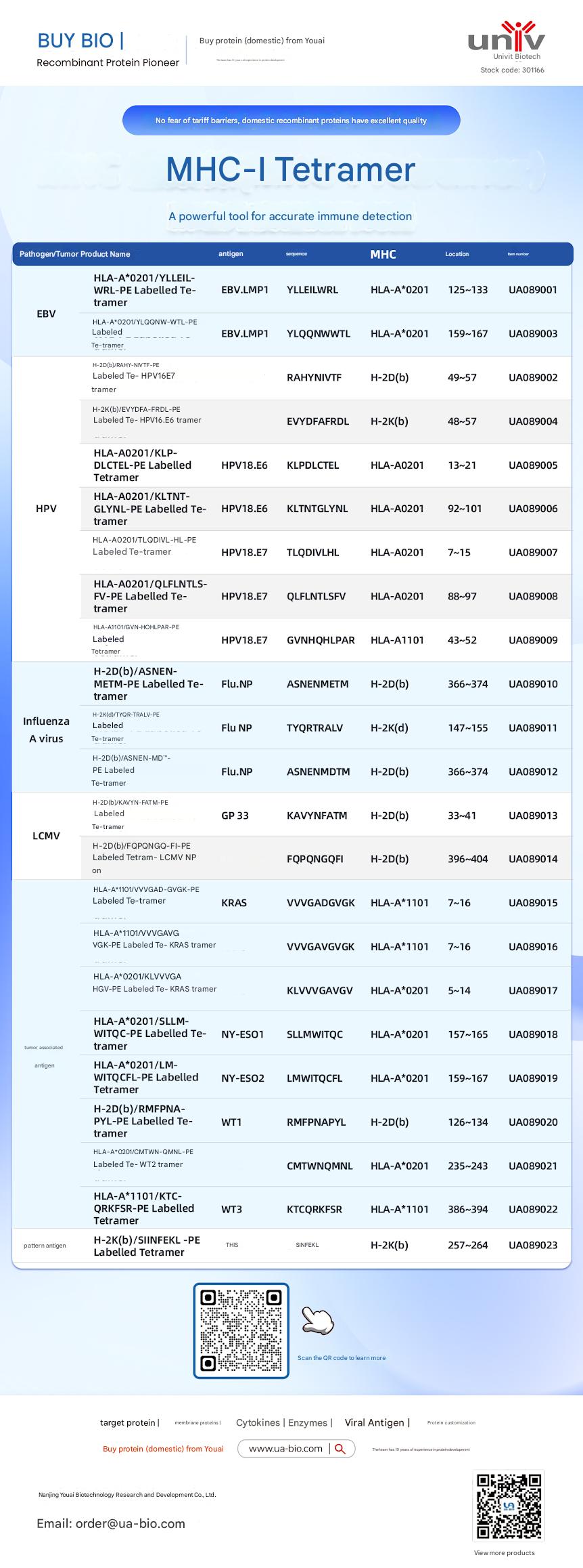
Related Products:
| Disease Category | Product Name | Antigen | Sequence | MHC | Position | Product Number |
|---|---|---|---|---|---|---|
| EBV | HLA-A*0201/YLELLVWRL-PE Labelled Tetramer | EBV.LMP1 | YLELLVWRL | HLA-A*0201 | 125-133 | UA089001 |
| EBV | HLA-A*0201/YLQQNWTL-PE Labelled Tetramer | EBV.LMP1 | YLQQNWTL | HLA-A*0201 | 159-167 | UA089003 |
| EBV | H-2Db(b)/RAHY-NIVTF-PE Labelled Tetramer | HPV16.E7 | RAHYNIVTF | H-2Db | 49-57 | UA089002 |
| HPV | H-2K(b)/EVYDFA-FRQL-PE Labelled Tetramer | HPV16.E6 | EVYDFARDL | H-2Kb | 48-57 | UA089004 |
| HPV | HLA-A*0201/KLP-DLCTL-PE Labelled Tetramer | HPV18.E6 | KLPDCTL | HLA-A*0201 | 13-21 | UA089005 |
| HPV | HLA-A*0201/KLTNT-GLYQL-PE Labelled Tetramer | HPV18.E6 | KLTNTGLYNL | HLA-A*0201 | 92-101 | UA089006 |
| HPV | HLA-A*0201/TLODIVIHL-PE Labelled Tetramer | HPV18.E7 | TLODIVIHL | HLA-A*0201 | 7~15 | UA089007 |
| HPV | HLA-A*0201/QFLNTL-FV-PE Labelled Tetramer | HPV18.E7 | QFLNTLFSV | HLA-A*0201 | 88-97 | UA089008 |
| HPV | HLA-A*1101/GVNHQLPAR-PE Labelled Tetramer | HPV18.E7 | GVNHQLPAR | HLA-A*1101 | 43-52 | UA089009 |
| Influenza A Virus | H-2D(b)/ASNENMETM-PE Labelled Tetramer | Flu.NP | ASNENMETM | H-2Db | 366-374 | UA089010 |
| Influenza A Virus | H-2K(d)/TYQR-TRALY-PE Labelled Tetramer | Flu.NP | TYQRTRALY | H-2Kd | 147-155 | UA089011 |
| Influenza A Virus | H-2D(b)/ASNEN-MDTM-PE Labelled Tetramer | Flu.NP | ASNENMDTM | H-2Db | 366-374 | UA089012 |
| LCMV | H-2D(b)/KAVYNFATM-PE Labelled Tetramer | GP 33 | KAVYNFATM | H-2Db | 33-41 | UA089013 |
| LCMV | H-2D(b)/FQPGQGFVK-PE Labelled Tetramer | LCMV NP | FQPGQGFVK | H-2Db | 396-404 | UA089014 |
| Tumor-related | HLA-A*1101/VVGADGVK-PE Labelled Tetramer | KRAS | VVGADGVK | HLA-A*1101 | 7~16 | UA089015 |
| Tumor-related | HLA-A*1101/VVGAGVGK-PE Labelled Tetramer | KRAS | VVGAGVGK | HLA-A*1101 | 7~16 | UA089016 |
| Tumor-related | HLA-A*0201/KLVVGAGV-PE Labelled Tetramer | KRAS | KLVVGAGV | HLA-A*0201 | 5~14 | UA089017 |
| Tumor-related | HLA-A*0201/SLLMWITQC-PE Labelled Tetramer | NY-ESO1 | SLLMWITQC | HLA-A*0201 | 157-165 | UA089018 |
| Melanoma | HLA-A*0201/LMWITQCFL-PE Labelled Tetramer | NY-ESO2 | LMWITQCFL | HLA-A*0201 | 159-167 | UA089019 |
| Melanoma | H-2Db(b)/MMFPNA-P1-PE Labelled Tetramer | WT1 | RMFPNAPL | H-2Db | 126-134 | UA089020 |
| Melanoma | HLA-A*0201/CMTWV-PE Labelled Tetramer | WT2 | CMTWVNMDM | HLA-A*0201 | 235-243 | UA089021 |
| Melanoma | HLA-A*1101/KTCQRKSF-PE Labelled Tetramer | WT3 | KTCQRKSF | HLA-A*1101 | 386-394 | UA089022 |
| Ovarian Cancer | H-2K(b)/SINFEKL-PE Labelled Tetramer | OVA | SINFEKL | H-2Kb | 257-264 | UA089023 |
Product Information