MHC
On November 27, 2019, the American Association of Immunologists' official journal, The Journal of Immunology (JI), published online the research by Professor Xia Chun's team from China Agricultural University, including doctoral student Ma Lizhen. The study first determined the crystal structure of the African clawed frog (Xenopus laevis) MHC-I molecule with an antigenic peptide, uncovering unique conformational features of antigen presentation in amphibians and the structural basis of their defense against iridovirus infection.
The team's high - resolution crystallographic analysis revealed significant species - specific conformational changes in the African clawed frog's pMHC - I antigen - binding groove. A unique glutamic acid - valine dipeptide insertion mutation in the α - helix connecting region of the α2 domain was identified (Figure 1A, red box). This mutation causes the antigen - binding groove of the African clawed frog's pMHC - I to differ markedly from that of other jawed vertebrates. Structural comparison showed that the insertion sequence raises the highest point on the surface of the African clawed frog's pMHC - I molecule by about 3.8 Å compared to other species (Figure 1B). Although the mutation site doesn't directly participate in the contact interface between pMHC - I and TCR (Figure 1C, red area), molecular dynamics simulations and surface plasmon resonance assays confirmed that the steric hindrance change can induce specific TCR docking mode reconfiguration (Figure 1D), forming a unique interaction paradigm of pMHC - I - TCR in amphibians. The study also systematically analyzed the binding spectrum characteristics of African clawed frog pMHC - I molecules with iridovirus - derived antigenic peptides using novel peptide mass spectrometry, providing structural evidence for understanding amphibian antiviral immunity.

Figure 1. Unique conformation of Xenopus pMHC - I and its docking pattern with T cell receptors. A. Overall conformation of the Xenopus pMHC - I complex, with the inserted glutamic acid - valine dipeptide shown in stick form and the blue mesh representing the 2Fo - Fc electron density map (at the 1.5σ level); B. Structural overlay of the antigen - binding groove of Xenopus pMHC - I (green cartoon model) with other species (white), with the red dashed box highlighting the conformational difference caused by the insertion sequence; C. Close - up view of the antigen - binding groove, with the red semi - transparent surface indicating the TCR contact interface and the yellow stick model representing the Xenopus - specific inserted amino acids; D. Docking pattern of Xenopus pMHC - I (green) with human TCR (cyan), with the red circle highlighting the steric hindrance effect caused by the inserted amino acids.
As a key transitional group from aquatic to terrestrial life, the structural analysis of Xenopus pMHC - I provides crucial molecular evidence for understanding vertebrate immune system evolution. Phylogenetic comparative analysis revealed that in non - mammalian species (such as Xenopus, chickens, and grass carp), the AB loop of pMHC - I molecules directly contacts the light chain β2 - microglobulin (β2m) (Figures 2A - C), whereas in mammals, this domain exhibits an independent conformation (Figure 2D). Furthermore, spatial distance measurements indicated that during vertebrate evolution from fish to mammals, the vertical distance from key amino acid residues in the pMHC - I - CD8 coreceptor interaction interface to the CD8 molecular surface gradually decreased (fish: 18.4 ± 2.1 Å; amphibians: 15.7 ± 1.9 Å; birds: 12.3 ± 1.6 Å; mammals: 9.1 ± 1.2 Å). This suggests that after vertebrates adapted to terrestrial life, the molecular recognition accuracy and binding affinity between pMHC - I and CD8 auxiliary receptors significantly increased. This structure - function - coupled evolutionary pattern offers a structural biology perspective for explaining the molecular driving mechanisms of adaptive immune systems in species radiation adaptation.
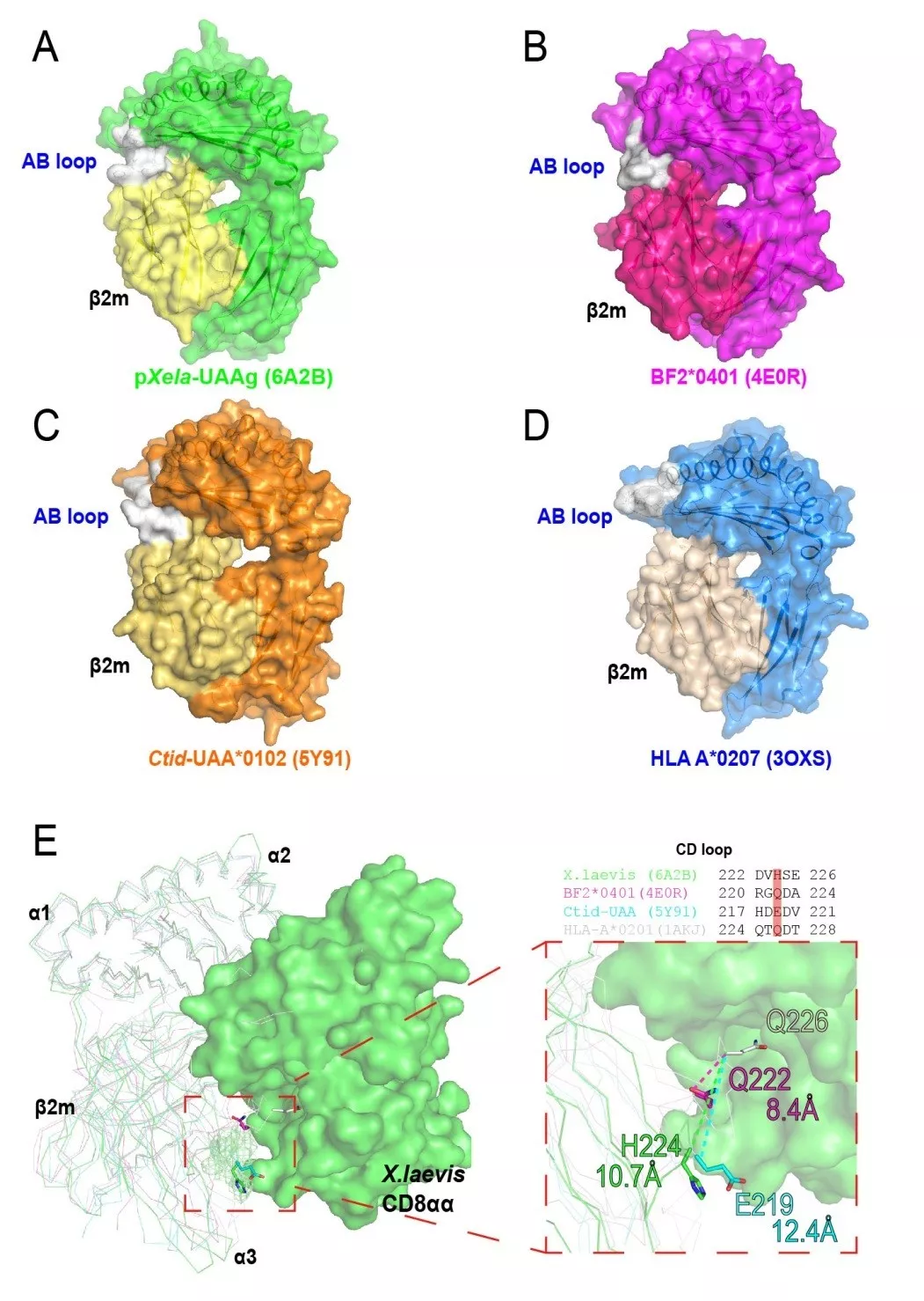
Figure 2. Structural features of amphibian pMHC - I in evolution. A. Structure of the amphibian pMHC - I complex, with the gray area indicating the contact interface between the AB loop of the antigen - binding groove and β2m; B. Structure of avian pMHC - I, with the gray area showing the direct interaction between the AB loop and the light chain; C. Structure of piscine pMHC - I, with the gray area highlighting the AB loop - β2m contact interface; D. Structure of mammalian pMHC - I, with the gray area showing the independent conformation of the AB loop; E. Cross - species structural comparison: superposition of pMHC - I molecules from fish (cyan), amphibians (green), birds (purple), and mammals (white), with a cartoon model of the homology - modeled amphibian CD8 molecule presented and key interacting amino acids indicated by colored stick models (red: fish; orange: amphibians; yellow: birds; blue: mammals).
The African clawed frog, a classic model organism, is indispensable in immunological and developmental biological research. Its unique metamorphic development process is accompanied by immune system reprogramming: MHC - I expression is restricted in the tadpole stage but widely expressed in adult frogs. This stage - specific immunological feature is closely related to viral infection susceptibility. Frog virus 3 (FV3) is lethal to tadpoles but can be effectively cleared in adult frogs. To explore the antiviral immune mechanism, this study used in - vitro - refolding technology with a random nonapeptide library to systematically identify the antigenic peptide spectrum of Xenopus MHC - I molecules (Figure 3) and performed high - throughput antigenic epitope scanning of the FV3 virome. Results showed that 689 nonapeptide sequences with high - affinity binding to Xenopus MHC - I were identified from 87 FV3 - encoded proteins. Combined with immunohistochemical analysis, these viral peptides were found to induce specific CD8⁺ T cell responses in adult frog spleens, with cytotoxic activity increasing by about 4.7 - fold compared to the tadpole stage. Furthermore, functional validation demonstrated that dendritic cells pulsed with MHC - I - restricted viral peptides significantly enhanced T cell degranulation (CD107a expression increased from 12.4% to 38.7%) and IFN - γ secretion levels (ELISPOT counts rose from 82 ± 15 to 294 ± 37) in adult frogs. These data support the hypothesis that enhanced MHC - I expression post - metamorphosis activates specific cytotoxic T lymphocyte (CTL) responses, forming a key immune defense against FV3 infection in adult frogs.
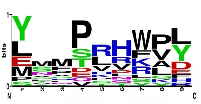
Figure 3. Antigenic peptide spectrum of Xenopus MHC - I molecules. This figure presents the heat map generated by calculating the weighted probabilities of amino acids at each peptide position based on the mass spectrometry analysis of the in - vitro random nonapeptide library binding experiment, systematically decoding the antigenic peptide - binding preferences of Xenopus MHC - I molecules.
The of the fine structure of the Xenopus pMHC - I complex holds dual scientific value. In evolutionary immunology, it first fully reveals the three - dimensional conformational characteristics of amphibian MHC - I molecules, filling a key gap in the evolutionary lineage of the vertebrate adaptive immune system. Regarding functional mechanisms, the systematic analysis of antigen - peptide binding spectra clarifies the molecular basis of antigen - peptide recognition and presentation by Xenopus MHC - I molecules. The peptide - binding groove shows a marked preference for hydrophobic amino acids, especially C - terminal Val/Leu, directly linked to FV3 virus - specific CTL responses in adult frogs. This achievement provides structural evidence for understanding immune evolution in amphibians during the transition from aquatic to terrestrial environments and lays a foundation for designing novel MHC - I - restricted epitope - based antiviral vaccines through high - throughput screening of viral antigenic epitopes.
Related Products:
| Disease Category |
Product Name |
Antigen |
Sequence |
MHC |
Position |
Product Number |
| EBV |
HLA-A*0201/YLELLVWRL-PE Labelled Tetramer |
EBV.LMP1 |
YLELLVWRL |
HLA-A*0201 |
125-133 |
UA089001 |
| EBV |
HLA-A*0201/YLQQNWTL-PE Labelled Tetramer |
EBV.LMP1 |
YLQQNWTL |
HLA-A*0201 |
159-167 |
UA089003 |
| EBV |
H-2Db(b)/RAHY-NIVTF-PE Labelled Tetramer |
HPV16.E7 |
RAHYNIVTF |
H-2Db |
49-57 |
UA089002 |
| HPV |
H-2K(b)/EVYDFA-FRQL-PE Labelled Tetramer |
HPV16.E6 |
EVYDFARDL |
H-2Kb |
48-57 |
UA089004 |
| HPV |
HLA-A*0201/KLP-DLCTL-PE Labelled Tetramer |
HPV18.E6 |
KLPDCTL |
HLA-A*0201 |
13-21 |
UA089005 |
| HPV |
HLA-A*0201/KLTNT-GLYQL-PE Labelled Tetramer |
HPV18.E6 |
KLTNTGLYNL |
HLA-A*0201 |
92-101 |
UA089006 |
| HPV |
HLA-A*0201/TLODIVIHL-PE Labelled Tetramer |
HPV18.E7 |
TLODIVIHL |
HLA-A*0201 |
7~15 |
UA089007 |
| HPV |
HLA-A*0201/QFLNTL-FV-PE Labelled Tetramer |
HPV18.E7 |
QFLNTLFSV |
HLA-A*0201 |
88-97 |
UA089008 |
| HPV |
HLA-A*1101/GVNHQLPAR-PE Labelled Tetramer |
HPV18.E7 |
GVNHQLPAR |
HLA-A*1101 |
43-52 |
UA089009 |
| Influenza A Virus |
H-2D(b)/ASNENMETM-PE Labelled Tetramer |
Flu.NP |
ASNENMETM |
H-2Db |
366-374 |
UA089010 |
| Influenza A Virus |
H-2K(d)/TYQR-TRALY-PE Labelled Tetramer |
Flu.NP |
TYQRTRALY |
H-2Kd |
147-155 |
UA089011 |
| Influenza A Virus |
H-2D(b)/ASNEN-MDTM-PE Labelled Tetramer |
Flu.NP |
ASNENMDTM |
H-2Db |
366-374 |
UA089012 |
| LCMV |
H-2D(b)/KAVYNFATM-PE Labelled Tetramer |
GP 33 |
KAVYNFATM |
H-2Db |
33-41 |
UA089013 |
| LCMV |
H-2D(b)/FQPGQGFVK-PE Labelled Tetramer |
LCMV NP |
FQPGQGFVK |
H-2Db |
396-404 |
UA089014 |
| Tumor-related |
HLA-A*1101/VVGADGVK-PE Labelled Tetramer |
KRAS |
VVGADGVK |
HLA-A*1101 |
7~16 |
UA089015 |
| Tumor-related |
HLA-A*1101/VVGAGVGK-PE Labelled Tetramer |
KRAS |
VVGAGVGK |
HLA-A*1101 |
7~16 |
UA089016 |
| Tumor-related |
HLA-A*0201/KLVVGAGV-PE Labelled Tetramer |
KRAS |
KLVVGAGV |
HLA-A*0201 |
5~14 |
UA089017 |
| Tumor-related |
HLA-A*0201/SLLMWITQC-PE Labelled Tetramer |
NY-ESO1 |
SLLMWITQC |
HLA-A*0201 |
157-165 |
UA089018 |
| Melanoma |
HLA-A*0201/LMWITQCFL-PE Labelled Tetramer |
NY-ESO2 |
LMWITQCFL |
HLA-A*0201 |
159-167 |
UA089019 |
| Melanoma |
H-2Db(b)/MMFPNA-P1-PE Labelled Tetramer |
WT1 |
RMFPNAPL |
H-2Db |
126-134 |
UA089020 |
| Melanoma |
HLA-A*0201/CMTWV-PE Labelled Tetramer |
WT2 |
CMTWVNMDM |
HLA-A*0201 |
235-243 |
UA089021 |
| Melanoma |
HLA-A*1101/KTCQRKSF-PE Labelled Tetramer |
WT3 |
KTCQRKSF |
HLA-A*1101 |
386-394 |
UA089022 |
| Ovarian Cancer |
H-2K(b)/SINFEKL-PE Labelled Tetramer |
OVA |
SINFEKL |
H-2Kb |
257-264 |
UA089023 |












 Business license (three certificates in one)