




MHC-IIb Tetramers: Decoding New Dimensions of Immune Tolerance and Antitumor Responses Regulated by Noncanonical MHC Molecules.


MHC class I and class II molecules, via sophisticated antigen processing and presentation pathways, respectively drive the activation of cytotoxic T lymphocytes (CTLs) and helper T cells (Th), serving as the initiation point for adaptive immune responses.
- Recent Advances
- Product Information
MHC-IIb Tetramers
In the study of tumor immune responses, T cell cytotoxicity, and antigen-presentation mechanisms, the major histocompatibility complex (MHC) serves as the "central molecular identification system of the immune system," with its functional elucidation holding foundational significance. MHC class I and II molecules establish sophisticated antigen-processing pathways that govern the activation of cytotoxic T lymphocytes (CTLs) and helper T cells (Th), respectively, collectively forming the initiation nodes of adaptive immune responses. However, tumor cells can evade immune detection by modulating MHC expression profiles through multiple mechanisms, making this molecular interplay network a critical target for breakthroughs in current tumor immunotherapy strategies.
Dual Roles and Immunoregulatory Mechanisms of MHC Molecules
MHC class I molecules are ubiquitously expressed on nearly all nucleated cell surfaces, activating CD8⁺ CTLs by presenting endogenous antigenic peptides (e.g., viral proteins or tumor-mutation-associated antigens), thereby forming a systemic immune surveillance network. Downregulation of their expression or defects in antigen-processing pathways (e.g., TAP transporter dysfunction) directly enable tumor cells to evade CTL-mediated killing—a phenomenon supported by pathological evidence in melanomas, lung cancers, and other solid tumors. In contrast, MHC class II expression is strictly confined to professional antigen-presenting cells (APCs), activating CD4⁺ Th cells through exogenous antigen processing to coordinate humoral and cellular immune responses. Notably, regulatory T cells (Tregs) and myeloid-derived suppressor cells (MDSCs) within the tumor microenvironment can secrete immunosuppressive cytokines (e.g., IL-10, TGF-β) to downregulate MHC class II expression on APCs, thereby blocking the antitumor immune cascade.
Aberrant MHC Regulation in Tumor Immune Evasion
Tumor cells evolve multiple strategies to disrupt MHC function:
- MHC Class I Downregulation: Through epigenetic silencing (e.g., HLA-I gene promoter methylation) or interference with β2-microglobulin synthesis, tumors evade CTL recognition—a mechanism linked to primary resistance against PD-1/PD-L1 inhibitors.
- Aberrant Antigen-Presentation Pathways: Some tumors upregulate non-classical MHC-Ib molecules (e.g., HLA-E) to substitute classical MHC-Ia molecules, engaging inhibitory receptors (e.g., NKG2A) and suppressing NK cell activity.
- MHC Class II Dysregulation: Tumor-associated macrophages (TAMs) can induce abnormal MHC class II expression coupled with loss of costimulatory molecules (e.g., CD80/CD86), creating an "ineffective antigen-presentation" phenotype that induces T cell exhaustion.
Therapeutic Strategies Targeting MHC Molecules and Challenges
Immunotherapeutic interventions targeting MHC molecules are emerging as research hotspots:
- MHC Class I Restoration: Histone deacetylase inhibitors (HDACis) can upregulate tumor MHC-I expression via epigenetic modulation, enhancing CTL recognition.
- MHC Class II-Targeted Vaccines: Personalized vaccines based on tumor-specific neoantigens rely on efficient APC uptake and presentation of MHC class II-restricted antigens.
- Non-Classical MHC Blockade: Monoclonal antibodies targeting the HLA-E/NKG2A axis (e.g., Monalizumab) are under clinical investigation to relieve NK cell suppression and restore immune surveillance.
The MHC system, bridging innate and adaptive immunity, profoundly influences tumor microenvironment remodeling through its regulatory network. Deepening the understanding of MHC-I/II dual regulation will provide critical molecular targets for developing next-generation immunotherapies.
1. Background: What Are MHC Molecules?
MHC (Major Histocompatibility Complex) represents a family of highly polymorphic cell-surface glycoproteins with core biological functions in:
- Antigen Presentation and Immune Recognition: By specifically binding antigenic peptides, MHC molecules form the molecular interface for T cell receptor (TCR) recognition, initiating and regulating adaptive immune responses.
In humans, the MHC is termed the HLA (Human Leukocyte Antigen) system, located on the short arm of chromosome 6. Serving as the immune system's "molecular ID," HLA molecules, expressed on nearly all nucleated cells, form the foundational framework for T cells to distinguish self from non-self antigens.
MHC molecules are categorized into two classes based on distribution and function:
| Class | Primary Role | Activation Target |
|---|---|---|
| MHC-I | Present endogenous antigens (e.g., viral proteins, tumor-mutation-associated antigens) to cytotoxic T cells (CD8⁺ T cells) | CD8⁺ T cells (Cytotoxic T lymphocytes) |
| MHC-II | Present exogenous antigens (e.g., extracellular bacterial proteins, fungal polysaccharides) to helper T cells (CD4⁺ T cells) | CD4⁺ T cells (Helper T lymphocytes) |
Functional Analysis:
- MHC-I Molecules: Process cytoplasmic proteins via the ubiquitin-proteasome system, presenting 8-11 amino acid peptides to CD8⁺ T cells to trigger direct killing of infected or tumor cells. Universal expression covers all nucleated cells.
- MHC-II Molecules: Degrade exogenous antigens through endosomal-lysosomal pathways, generating 13-25 amino acid peptides to activate CD4⁺ T cells, thereby coordinating immune responses (e.g., B cell antibody production, macrophage activation). Expression is strictly limited to professional APCs like dendritic cells, B lymphocytes, and macrophages.
This division ensures comprehensive monitoring of intracellular abnormalities (MHC-I pathway) and precise detection of environmental threats (MHC-II pathway), forming the core architecture of multi-layered immune defense.
2. Classic MHC Molecules at a Glance (Human HLA System)
MHC-I Molecules (Ubiquitously Expressed on All Nucleated Cell Surfaces)
| Molecule | Functional Analysis |
|---|---|
| HLA-A | Core endogenous antigen presenter, specializing in tumor neoantigens and viral peptides (e.g., HPV E7). Its α1/α2 domain forms a peptide-binding groove stabilized by anchor residues and C-terminal hydrophobic amino acids. |
| HLA-B | The most polymorphic MHC-I subclass in humans. Its highly variable alleles (e.g., B07:02, B08:01) provide the TCR recognition diversity critical for eliminating intracellular pathogens (e.g., Mycobacterium tuberculosis). |
| HLA-C | Combines classical MHC-I functions with immunoregulatory roles: modulates NK cell activity via killer immunoglobulin-like receptors (KIRs). Low expression levels play special roles in pregnancy tolerance and HIV progression. |
Structural Features: MHC-I molecules are heterodimers of a heavy chain (α chain) and β2-microglobulin. The α3 domain binds CD8, ensuring precise TCR signaling, while β2-microglobulin stabilizes α chain conformation to maintain antigen-presentation integrity.
MHC-II Molecules (Exclusively Expressed on APC Surfaces)
| Molecule | Functional Analysis |
|---|---|
| HLA-DR | Central hub for immune regulation. DRB1 allele polymorphism directly affects autoimmune disease susceptibility (e.g., rheumatoid arthritis-associated DRB1*04:01) and modulates CD4⁺ T cell differentiation (Th1/Th2/Th17). |
| HLA-DP | Mediates specific antigen recognition and T cell activation. Its DPA1/DPB1 heterodimer selectively binds peptides containing proline or basic amino acids via unique α1/β1 domain conformations, promoting B cell antibody class switching during humoral responses. |
| HLA-DQ | Key autoimmune susceptibility gene. The DQA105:01/DQB102:01 haplotype is strongly linked to celiac disease. Aberrant expression disrupts Treg tolerance, leading to autoreactive T cell activation. |
Structural Features: MHC-II molecules are α/β heterodimers with an open peptide-binding groove (α2/β2 domains) accommodating 13-25 amino acid peptides. Their expression is induced by cytokines like IFN-γ, amplifying immune activation.
Functional Extensions:
- MHC-I/II Synergy: During cross-presentation, MHC-I molecules can transiently express on APCs to present exogenous antigens to CD8⁺ T cells, while MHC-II molecules may aberrantly present endogenous peptides, contributing to autoimmune pathogenesis.
- Epigenetic Regulation: Tumors often silence MHC expression via DNA methylation (e.g., HLA-I promoter CpG island hypermethylation) or histone modifications (e.g., H3K27me3), enabling immune evasion.
3. Antigen-Presentation Mechanisms: How Do MHC Molecules "Show" Antigens to the Immune System?
MHC-I Pathway: Endogenous Antigen Processing
- Cytosolic proteins (e.g., viral, tumor-derived) are cleaved into 8-11 amino acid peptides by the ubiquitin-proteasome system.
- The antigen-processing transporter (TAP) pumps peptides into the ER lumen.
- Chaperones (e.g., calreticulin) assist peptide-MHC-I complex formation, with β2-microglobulin stabilizing the α chain.
- Mature MHC-I-peptide complexes traffic to the cell membrane for TCR recognition by CD8⁺ T cells, triggering cytotoxicity (e.g., perforin/granzyme release or FasL-induced apoptosis).
MHC-II Pathway: Exogenous Antigen Cross-Presentation
- APCs internalize exogenous antigens via phagocytosis or receptor-mediated endocytosis, forming phagosomes.
- Acidic hydrolases (e.g., cathepsins B/D/L) degrade proteins into 13-25 amino acid peptides in phagolysosomes.
- Newly synthesized MHC-II molecules bind the invariant chain (Ii), whose CLIP fragment occupies the peptide-binding groove to prevent nonspecific peptide binding.
- The LLN motif targets MHC-II-Ii complexes to endolysosomes, where cathepsin S removes Ii, leaving CLIP in the groove until HLA-DM facilitates high-affinity peptide exchange.
- Mature MHC-II-peptide complexes reach the cell membrane, activating CD4⁺ T cells via TCR and costimulatory signals (e.g., CD80/CD86), amplifying immune responses (e.g., Th1 cytokine secretion, B cell antibody class switching, memory T cell generation).
Synergistic Mechanisms and Immune Regulation
- Cross-Presentation: Some APCs (e.g., CD8α⁺ dendritic cells) present exogenous antigens via MHC-I, bridging innate and adaptive immunity.
- Epitope Spreading: MHC-II-presented peptides induce CD4⁺ T cells to secrete IFN-γ, upregulating MHC-I and enhancing CTL activity.
- Checkpoint Regulation: Tumors exploit MHC-I downregulation or PD-L1 expression to create an "antigen-presentation defect-immune suppression" loop, a key target for immunotherapy.

4. MHC and Tumor Immune Evasion
Tumors employ multiple molecular strategies to evade immunity, with MHC dysregulation playing a central role:
- MHC-I Loss: Epigenetic silencing (e.g., HLA-I promoter hypermethylation), β2-microglobulin (B2M) mutations, or miRNA regulation (e.g., miR-200 targeting TAP) impair CTL recognition, causing "immune ignorance."
- Antigen-Processing Blockade: Inhibiting proteasome components (e.g., PSMB8 mutations) or TAP1/TAP2 disrupts peptide transport, leading to "antigen-presentation defects."
- MHC-II Suppression: Silencing MHC-II transactivator (CIITA) or interferon regulatory factor 1 (IRF1) reduces CD4⁺ T cell help, promoting Treg accumulation and Th1 imbalance.
- Non-Classical MHC Upregulation: Some tumors express non-classical MHC-Ib molecules (e.g., HLA-E/G) to engage inhibitory receptors (e.g., NKG2A/CD94), suppressing NK cells and enabling "checkpoint evasion."
These mechanisms allow tumors to evade detection despite high mutational burdens or immunogenic neoantigens, crippling immune surveillance.
5. Research and Applications: Why MHC Molecules Matter
In tumor immunotherapy, MHC expression profiles are critical for predicting efficacy and designing interventions:
- Therapeutic Efficacy Prediction:
- MHC-I loss correlates with primary resistance to PD-1/PD-L1 inhibitors, with ~30% of patients showing HLA-I LOH or B2M mutations.
- High MHC-II expression (e.g., HLA-DRB1*04:01) associates with increased TIL density, PD-L1 expression, and prolonged survival, identifying immunotherapy-responsive populations.
- Emerging Interventions:
- Epigenetic Modulators: HDACis reverse HLA-I promoter hypermethylation, restoring CTL sensitivity.
- Gene Therapy: CIITA adenovirus vectors induce tumor MHC-II expression, reshaping the microenvironment.
- Personalized Vaccines: Neoantigen-mRNA vaccines (e.g., Moderna's mRNA-4157) and peptide vaccines (e.g., NeoVax) show efficacy in melanoma and glioblastoma.
- Biomarker Development: Multidimensional models integrating MHC expression, antigen-processing mutations (e.g., TAP1/PSMB8), and TCR diversity (e.g., Immunophenoscore) guide treatment decisions.
Conclusion: The Importance of Understanding MHC Molecules
As the immune system's "molecular ID" and "danger signal platform," MHC expression networks determine whether tumors are detected and eliminated. In the era of personalized immunotherapy, deepening MHC-I/II regulation insights is key to understanding tumor evasion and developing next-gen therapies (e.g., bispecific antibodies, TCR-engineered T cells). Precision interventions targeting MHC molecules promise to reshape tumor microenvironment balance, advancing immunotherapy into a "molecular subtyping-driven" paradigm.
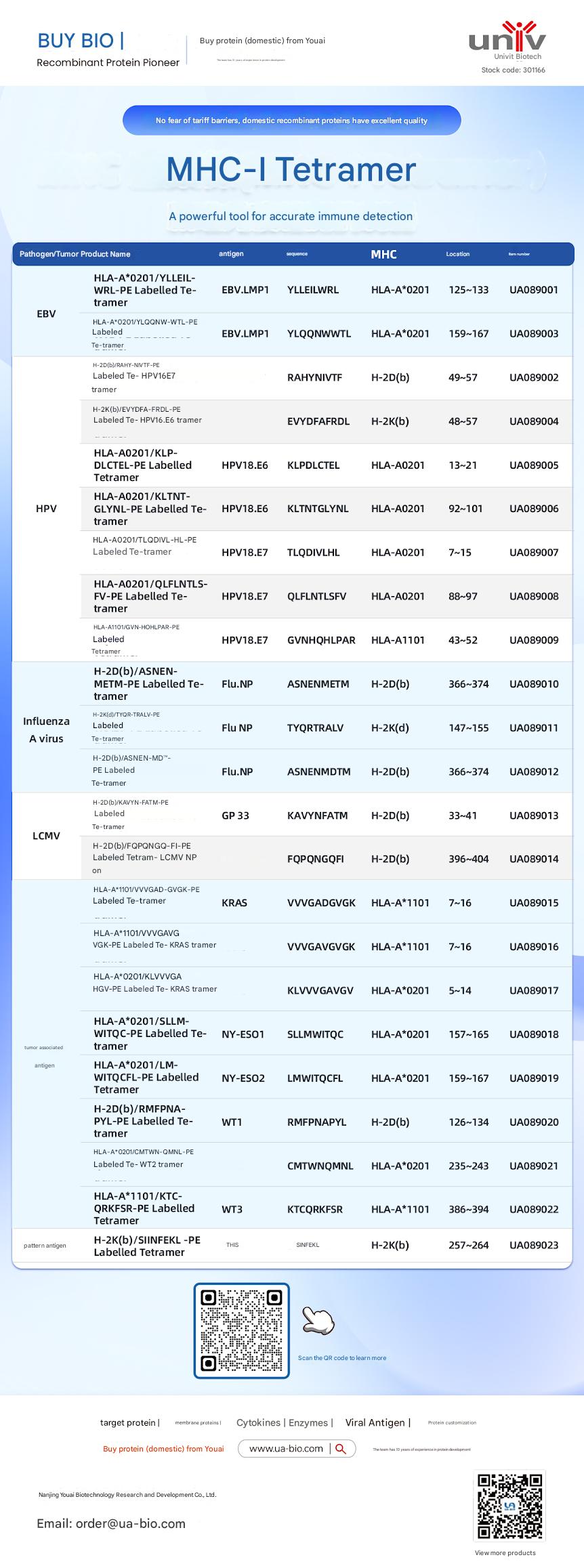
| Disease Category | Product Name | Antigen | Sequence | MHC | Position | Product Number |
|---|---|---|---|---|---|---|
| EBV | HLA-A*0201/YLELLVWRL-PE Labelled Tetramer | EBV.LMP1 | YLELLVWRL | HLA-A*0201 | 125-133 | UA089001 |
| EBV | HLA-A*0201/YLQQNWTL-PE Labelled Tetramer | EBV.LMP1 | YLQQNWTL | HLA-A*0201 | 159-167 | UA089003 |
| EBV | H-2Db(b)/RAHY-NIVTF-PE Labelled Tetramer | HPV16.E7 | RAHYNIVTF | H-2Db | 49-57 | UA089002 |
| HPV | H-2K(b)/EVYDFA-FRQL-PE Labelled Tetramer | HPV16.E6 | EVYDFARDL | H-2Kb | 48-57 | UA089004 |
| HPV | HLA-A*0201/KLP-DLCTL-PE Labelled Tetramer | HPV18.E6 | KLPDCTL | HLA-A*0201 | 13-21 | UA089005 |
| HPV | HLA-A*0201/KLTNT-GLYQL-PE Labelled Tetramer | HPV18.E6 | KLTNTGLYNL | HLA-A*0201 | 92-101 | UA089006 |
| HPV | HLA-A*0201/TLODIVIHL-PE Labelled Tetramer | HPV18.E7 | TLODIVIHL | HLA-A*0201 | 7~15 | UA089007 |
| HPV | HLA-A*0201/QFLNTL-FV-PE Labelled Tetramer | HPV18.E7 | QFLNTLFSV | HLA-A*0201 | 88-97 | UA089008 |
| HPV | HLA-A*1101/GVNHQLPAR-PE Labelled Tetramer | HPV18.E7 | GVNHQLPAR | HLA-A*1101 | 43-52 | UA089009 |
| Influenza A Virus | H-2D(b)/ASNENMETM-PE Labelled Tetramer | Flu.NP | ASNENMETM | H-2Db | 366-374 | UA089010 |
| Influenza A Virus | H-2K(d)/TYQR-TRALY-PE Labelled Tetramer | Flu.NP | TYQRTRALY | H-2Kd | 147-155 | UA089011 |
| Influenza A Virus | H-2D(b)/ASNEN-MDTM-PE Labelled Tetramer | Flu.NP | ASNENMDTM | H-2Db | 366-374 | UA089012 |
| LCMV | H-2D(b)/KAVYNFATM-PE Labelled Tetramer | GP 33 | KAVYNFATM | H-2Db | 33-41 | UA089013 |
| LCMV | H-2D(b)/FQPGQGFVK-PE Labelled Tetramer | LCMV NP | FQPGQGFVK | H-2Db | 396-404 | UA089014 |
| Tumor-related | HLA-A*1101/VVGADGVK-PE Labelled Tetramer | KRAS | VVGADGVK | HLA-A*1101 | 7~16 | UA089015 |
| Tumor-related | HLA-A*1101/VVGAGVGK-PE Labelled Tetramer | KRAS | VVGAGVGK | HLA-A*1101 | 7~16 | UA089016 |
| Tumor-related | HLA-A*0201/KLVVGAGV-PE Labelled Tetramer | KRAS | KLVVGAGV | HLA-A*0201 | 5~14 | UA089017 |
| Tumor-related | HLA-A*0201/SLLMWITQC-PE Labelled Tetramer | NY-ESO1 | SLLMWITQC | HLA-A*0201 | 157-165 | UA089018 |
| Melanoma | HLA-A*0201/LMWITQCFL-PE Labelled Tetramer | NY-ESO2 | LMWITQCFL | HLA-A*0201 | 159-167 | UA089019 |
| Melanoma | H-2Db(b)/MMFPNA-P1-PE Labelled Tetramer | WT1 | RMFPNAPL | H-2Db | 126-134 | UA089020 |
| Melanoma | HLA-A*0201/CMTWV-PE Labelled Tetramer | WT2 | CMTWVNMDM | HLA-A*0201 | 235-243 | UA089021 |
| Melanoma | HLA-A*1101/KTCQRKSF-PE Labelled Tetramer | WT3 | KTCQRKSF | HLA-A*1101 | 386-394 | UA089022 |
| Ovarian Cancer | H-2K(b)/SINFEKL-PE Labelled Tetramer | OVA | SINFEKL | H-2Kb | 257-264 | UA089023 |
This article is reviewed and published by the technical expert team of UA
Disclaimer: This article partially utilizes artificial intelligence assistance in its creation. If any content involves copyright or intellectual property issues, please let us know and we promise to verify and remove it as soon as possible.
Purchase recombinant protein, choose Nanjing UA-Bio
UA protein focuses on providing various protein reagents, raw materials, and services required for drug research and development, cell therapy, gene therapy, and basic scientific research, including drug target proteins, immune checkpoint proteins, cytokines, tool enzymes, customized protein expression, and full-length transmembrane protein development. Youai is committed to providing customers with high-quality products and professional services, and building a High-tech Biological Enterprise with International Competitiveness.







 Business license (three certificates in one)