




MHC-Ib Tetramers: Molecular Basis for Noncanonical Antigen Presentation and NK Cell Immunomodulation.


MHC class I molecules present endogenous antigen-derived peptides. They display short peptides (8 - 11 amino acids long) from the cytoplasm, like viral or tumor - related ones, to cytotoxic T lymphocytes (CTLs).
- Recent Advances
- Product Information
MHC-Ib Tetramers
MHC class I molecules showcase endogenously derived antigenic peptides through a specialized presentation mechanism, primarily displaying short peptides (8-11 amino acids in length) derived from cytoplasmic proteins—such as viral peptides associated with infections or tumor-associated antigens—to cytotoxic T lymphocytes (CTLs). This biological process involves three precisely regulated steps: first, the proteasome system generates antigenic peptides of appropriate length through ubiquitin-proteasome-mediated cleavage of abnormal proteins; second, the antigen-processing transporter (TAP) heterodimer actively translocates cytosolic peptides into the endoplasmic reticulum (ER) lumen; finally, with assistance from ER chaperones (e.g., calreticulin), antigenic peptides form stable complexes with the α1/α2 domains of MHC class I molecules, which are then transported to the cell membrane via the Golgi network, establishing the molecular basis for TCR recognition and immune surveillance.
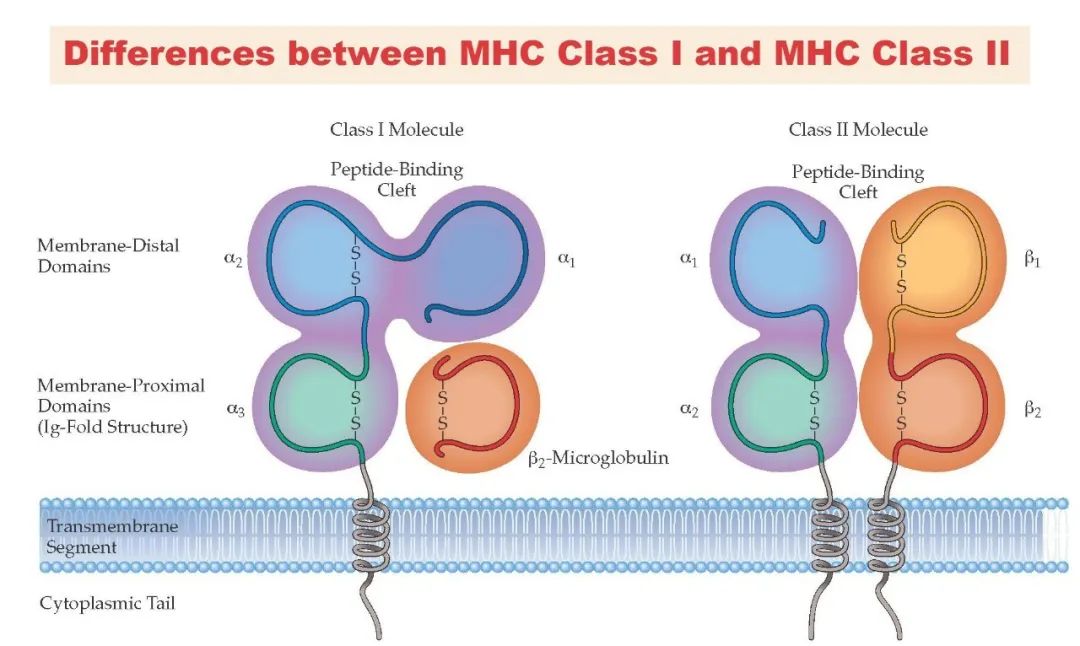
While both MHC class I and II molecules belong to the immunoglobulin superfamily and share antigen-presentation functions, they exhibit significant differences in antigen sources, cellular localization, and immune response regulation. After elucidating the mechanisms of MHC class I molecules, we now focus on the MHC class II system—these molecules exclusively process exogenous antigens (e.g., extracellular pathogen proteins), generating 13-25 amino acid-long peptides through endosomal-lysosomal pathways and specifically activating CD4⁺ T helper cells, thereby serving as central hubs in initiating and regulating adaptive immune responses.
MHC Class II Molecules
Encoded by genes in the HLA-D region of human chromosome 6, the MHC class II molecular system exhibits genetic polymorphisms analogous to MHC class I molecules—allelic variations between individuals directly result in differences in antigen-presentation repertoires. Notably, the peptide-binding groove of MHC class II molecules adopts a unique open conformation: unlike the closed ends of MHC class I binding clefts, MHC class II grooves remain open at both termini. This structural adaptation allows accommodation of extended antigenic peptides (13-25 amino acids), far exceeding the 8-11 amino acid capacity of MHC class I molecules. This structural-functional relationship enables MHC class II molecules to effectively present longer peptides derived from exogenous proteasomal processing, providing CD4⁺ T helper cells with a broader epitope-recognition interface.
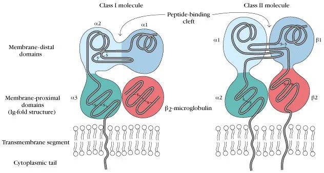
Before delving into MHC class II-mediated antigen presentation, we contrast the core functional differences between MHC class I and II molecules: MHC class I molecules recognize endogenous antigens (e.g., viral or tumor-derived proteins) via cross-presentation, delivering 8-11 amino acid short peptides to CTL TCRs; MHC class II molecules exclusively process exogenous antigens (e.g., extracellular bacterial or fungal proteins), generating 13-25 amino acid peptides through endosomal-lysosomal pathways to activate CD4⁺ T helper cells (Th cells).
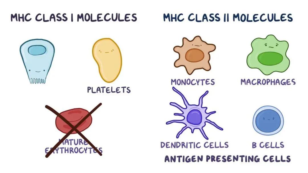
Cellular distribution analysis reveals that MHC class I molecules are constitutively expressed on nearly all nucleated cells, forming a systemic immune surveillance network. This widespread expression enables CTLs to continuously monitor protein synthesis across tissues, effectively containing viral infections or neoplastic transformations. In contrast, MHC class II expression is strictly confined to professional antigen-presenting cells (APCs)—including B lymphocytes, monocytes/macrophages, and dendritic cells—functioning as "environmental surveillance outposts." By selectively sampling local microenvironmental proteins, these cells amplify danger signals and relay them to the T cell response network. This differential expression strategy ensures comprehensive immune monitoring while preventing tissue damage from nonspecific activation.
MHC Class II-Mediated Antigen Presentation Process
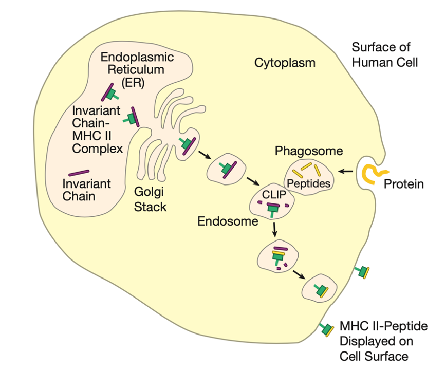
Following synthesis in the cytosol, MHC class II α and β chains are guided into the ER lumen via signal peptides. Within the ER, nascent MHC class II heterodimers specifically bind the invariant chain (Ii), a dedicated chaperone that regulates antigen presentation through:
- Endogenous Peptide Blocking: The Ii's CLIP (Class II-associated Invariant chain Peptide) fragment occupies the MHC class II binding groove, physically preventing nonspecific association with endogenous ER peptides (e.g., proteasome-generated short peptides). This segregation avoids competitive antigen loading between MHC class I and II molecules, ensuring dedicated roles in endogenous/exogenous antigen monitoring.
- Endosomal Targeting: The Ii's C-terminal endosomal sorting signal (e.g., LLN motif) directs MHC class II-Ii complexes to endosomal-lysosomal compartments via the Golgi network, bypassing conventional secretory pathways.
Within the acidic endosomal-lysosomal environment, the Ii undergoes progressive degradation: cathepsin S first cleaves most sequences, leaving only CLIP occupying the binding groove. Concurrently, exogenous antigen processing commences:
- Antigen Uptake and Processing: Cells internalize extracellular proteins via phagocytosis or receptor-mediated endocytosis, forming phagosomes. Fusion with endosomes enables acidic hydrolases (e.g., cathepsins B, L) to degrade intact proteins into 13-25 amino acid peptides.
- CLIP Displacement and Peptide Loading: HLA-DM (human)/H2-M (mouse) molecules act as dedicated editors, catalyzing CLIP release through conformational selection while stabilizing open MHC class II conformations to facilitate high-affinity peptide binding. This process is stringently filtered by peptide-binding motifs (e.g., HLA-DR anchor residue preferences).
Mature MHC class II-peptide complexes are finally transported to the cell membrane via vesicles, forming TCR-recognition and CD4 co-receptor-binding platforms.
Spatial Segregation Mechanisms of MHC Class I and II Antigen-Processing Systems
Evolution has refined antigen-presentation specialization through:
- Localization Isolation: MHC class I molecules load proteasome products directly in the ER, while MHC class II molecules process exogenous antigens in endosomal-lysosomal systems, establishing physical separation.
- Chaperone Regulation: The invariant chain and TAP (antigen peptide transporter) serve as exclusive chaperones for MHC class II and I molecules, respectively, ensuring antigen-loading specificity.
- Immune Surveillance Division: MHC class I molecules form a systemic surveillance network, while MHC class II molecules, through sampling by professional APCs, provide precise alerts against exogenous threats. This division ensures immune breadth while minimizing ineffective activation.
Summary
In this series of analyses, we have systematically elucidated the molecular characteristics and immunological functions of MHC class I and II molecules. Despite differences in subcellular localization, antigen preferences, and cellular distribution, both classes converge on antigen presentation—forming TCR-recognition interfaces through specific peptide binding. MHC class I molecules establish a systemic surveillance network via ubiquitous expression, detecting intracellular protein abnormalities in real time. MHC class II molecules, through sampling by professional APCs, precisely decode extracellular pathogen-associated molecular patterns. Together, these molecules constitute the initiation hub of adaptive immunity, with their peptide-binding grooves and membrane display forming the molecular foundation for T cell-mediated immune recognition. This complementary yet specialized antigen-presentation system represents a key mechanism for precise immune defense and homeostasis.
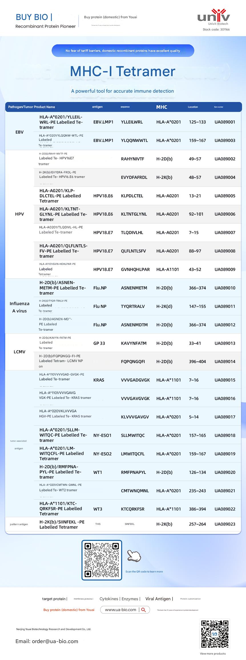
| Disease Category | Product Name | Antigen | Sequence | MHC | Position | Product Number |
|---|---|---|---|---|---|---|
| EBV | HLA-A*0201/YLELLVWRL-PE Labelled Tetramer | EBV.LMP1 | YLELLVWRL | HLA-A*0201 | 125-133 | UA089001 |
| EBV | HLA-A*0201/YLQQNWTL-PE Labelled Tetramer | EBV.LMP1 | YLQQNWTL | HLA-A*0201 | 159-167 | UA089003 |
| EBV | H-2Db(b)/RAHY-NIVTF-PE Labelled Tetramer | HPV16.E7 | RAHYNIVTF | H-2Db | 49-57 | UA089002 |
| HPV | H-2K(b)/EVYDFA-FRQL-PE Labelled Tetramer | HPV16.E6 | EVYDFARDL | H-2Kb | 48-57 | UA089004 |
| HPV | HLA-A*0201/KLP-DLCTL-PE Labelled Tetramer | HPV18.E6 | KLPDCTL | HLA-A*0201 | 13-21 | UA089005 |
| HPV | HLA-A*0201/KLTNT-GLYQL-PE Labelled Tetramer | HPV18.E6 | KLTNTGLYNL | HLA-A*0201 | 92-101 | UA089006 |
| HPV | HLA-A*0201/TLODIVIHL-PE Labelled Tetramer | HPV18.E7 | TLODIVIHL | HLA-A*0201 | 7~15 | UA089007 |
| HPV | HLA-A*0201/QFLNTL-FV-PE Labelled Tetramer | HPV18.E7 | QFLNTLFSV | HLA-A*0201 | 88-97 | UA089008 |
| HPV | HLA-A*1101/GVNHQLPAR-PE Labelled Tetramer | HPV18.E7 | GVNHQLPAR | HLA-A*1101 | 43-52 | UA089009 |
| Influenza A Virus | H-2D(b)/ASNENMETM-PE Labelled Tetramer | Flu.NP | ASNENMETM | H-2Db | 366-374 | UA089010 |
| Influenza A Virus | H-2K(d)/TYQR-TRALY-PE Labelled Tetramer | Flu.NP | TYQRTRALY | H-2Kd | 147-155 | UA089011 |
| Influenza A Virus | H-2D(b)/ASNEN-MDTM-PE Labelled Tetramer | Flu.NP | ASNENMDTM | H-2Db | 366-374 | UA089012 |
| LCMV | H-2D(b)/KAVYNFATM-PE Labelled Tetramer | GP 33 | KAVYNFATM | H-2Db | 33-41 | UA089013 |
| LCMV | H-2D(b)/FQPGQGFVK-PE Labelled Tetramer | LCMV NP | FQPGQGFVK | H-2Db | 396-404 | UA089014 |
| Tumor-related | HLA-A*1101/VVGADGVK-PE Labelled Tetramer | KRAS | VVGADGVK | HLA-A*1101 | 7~16 | UA089015 |
| Tumor-related | HLA-A*1101/VVGAGVGK-PE Labelled Tetramer | KRAS | VVGAGVGK | HLA-A*1101 | 7~16 | UA089016 |
| Tumor-related | HLA-A*0201/KLVVGAGV-PE Labelled Tetramer | KRAS | KLVVGAGV | HLA-A*0201 | 5~14 | UA089017 |
| Tumor-related | HLA-A*0201/SLLMWITQC-PE Labelled Tetramer | NY-ESO1 | SLLMWITQC | HLA-A*0201 | 157-165 | UA089018 |
| Melanoma | HLA-A*0201/LMWITQCFL-PE Labelled Tetramer | NY-ESO2 | LMWITQCFL | HLA-A*0201 | 159-167 | UA089019 |
| Melanoma | H-2Db(b)/MMFPNA-P1-PE Labelled Tetramer | WT1 | RMFPNAPL | H-2Db | 126-134 | UA089020 |
| Melanoma | HLA-A*0201/CMTWV-PE Labelled Tetramer | WT2 | CMTWVNMDM | HLA-A*0201 | 235-243 | UA089021 |
| Melanoma | HLA-A*1101/KTCQRKSF-PE Labelled Tetramer | WT3 | KTCQRKSF | HLA-A*1101 | 386-394 | UA089022 |
| Ovarian Cancer | H-2K(b)/SINFEKL-PE Labelled Tetramer | OVA | SINFEKL | H-2Kb | 257-264 | UA089023 |
This article is reviewed and published by the technical expert team of UA
Disclaimer: This article partially utilizes artificial intelligence assistance in its creation. If any content involves copyright or intellectual property issues, please let us know and we promise to verify and remove it as soon as possible.
Purchase recombinant protein, choose Nanjing UA-Bio
UA protein focuses on providing various protein reagents, raw materials, and services required for drug research and development, cell therapy, gene therapy, and basic scientific research, including drug target proteins, immune checkpoint proteins, cytokines, tool enzymes, customized protein expression, and full-length transmembrane protein development. Youai is committed to providing customers with high-quality products and professional services, and building a High-tech Biological Enterprise with International Competitiveness.







 Business license (three certificates in one)