




The Molecular Architecture, Genetic Polymorphism, and Immune Regulation Mechanisms of the Major Histocompatibility Complex (MHC)


The Major Histocompatibility Complex (MHC) is a highly polymorphic genetic system composed of a group of tightly linked genes. Its encoded products form the core molecular system for T lymphocyte recognition and antigen presentation. This antigen system is crucial for regulating allograft rejection in organ transplantation, with its molecular polymorphism directly impacting the specificity of immune responses.
- Recent Advances
- Product Information
MHC Antigens
The major histocompatibility complex (MHC) represents a highly polymorphic genetic system composed of a cluster of closely linked genes, whose products form the core molecular machinery mediating T lymphocyte recognition and antigen presentation. This antigen system serves as the key determinant regulating allograft rejection in transplantation, with its molecular polymorphism directly influencing the specificity of immune responses.
Based on molecular structure and functional characteristics, the MHC antigen system is categorized into two major classes: Class I and Class II antigens. Using the laboratory animal model mouse, the MHC antigen system is specifically termed the H-2 antigen system, with its gene cluster localized to chromosome 17. The murine H-2 complex encodes a complex array of products encompassing 11 functional subclasses:
Classical MHC Class I Molecules (MHC-Ia):
- H-2D Subclass: Encoded by H-2D genes, expressed on all nucleated cell surfaces
- H-2K Subclass: Includes alleles such as H-2Kk and H-2Kb
- H-2L Subclass: Exhibits allelic variations including H-2Ld and H-2Lq
Non-Classical MHC Class I Molecules (MHC-Ib):
- H-2Q Subclass: Demonstrates tissue-specific expression patterns
- H-2M Subclass: Involved in antigen processing and delivery
- H-2T Subclass: Functions in NK cell education
Classical MHC Class II Molecules (MHC-IIa):
- H-2A(I-A) Subclass: Composed of α and β chains forming heterodimers
- H-2E(I-E) Subclass: Exhibits cell-type-specific expression
- H-2P Subclass: Associated with immune regulation
Non-Classical MHC Class II Molecules (MHC-IIb):
- H-2M Subclass: Acts as a peptide editor during antigen loading
- H-2O Subclass: Expression regulated by cytokines
From a molecular conformation perspective, classical MHC Class I molecules consist of two polypeptide chains: a 45 kDa α chain (a transmembrane glycoprotein with extracellular domains α1, α2, and α3) and a 12 kDa β2-microglobulin (β2-MG) non-covalently associated with the α chain. The extracellular portion undergoes specific proteolytic cleavage by papain at the membrane-proximal 13th amino acid residue. Notably, human MHC Class I molecules (HLA-I) have α chain genes localized to the HLA-A, -B, and -C loci, while murine H-2K and H-2D/L genes form the core encoding components of Class I molecules.
MHC Class II molecules comprise 33 kDa α chains and 28 kDa β chains forming heterodimers through non-covalent interactions, with their antigen-binding groove formed by α1 and β1 domains. Both molecule classes exhibit conserved immunoglobulin-like fold domains in their membrane-proximal regions, justifying their classification within the immunoglobulin superfamily. This structural conservation correlates with their critical functions in mediating TCR-pMHC interactions and costimulatory signaling.
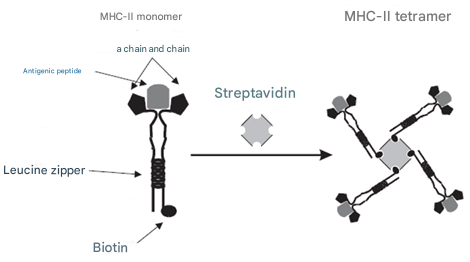
MHC Class I antigens display ubiquitous expression across nearly all nucleated cell membranes, primarily presenting endogenous antigenic information (including viral peptides from infections and tumor-associated antigens) to cytotoxic T lymphocytes (CTLs) via the α1/α2 domain-formed antigen-binding groove. This presentation mechanism specifically activates CD8⁺ T cells, triggering target cell lysis and viral clearance. In contrast, MHC Class II antigen expression is strictly lineage-restricted, primarily localized to professional antigen-presenting cells (including B lymphocytes, mononuclear phagocytes, dendritic cells, and epidermal Langerhans cells). Their biological function involves presenting exogenous antigens processed through endosomal pathways to CD4⁺ T helper cells via the α1/β1 heterodimeric groove, thereby initiating adaptive immune responses.
The MHC molecular system exhibits remarkable polymorphism following codominant haplotype inheritance patterns. In laboratory mouse models, inbred strains achieve genome-wide homozygosity through ≥20 consecutive generations of sibling mating, ensuring allele homogeneity at all H-2 loci. This genetic uniformity renders specific inbred strains ideal models for MHC functional studies, with expressed MHC haplotypes standardized using italicized letter codes (e.g., H-2^b, H-2^d). This nomenclature integrates all H-2 complex allele information, avoiding per-locus annotation complexity. For example, the BALB/c strain's complete MHC haplotype includes H-2K^d, H-2D^d, H-2L^d (Class I region) and I-A^d, I-E^d (Class II region), maintaining genetic integrity while enabling rapid phenotypic identification across strains.
As MHC alleles follow codominant inheritance, heterozygous individuals concurrently express parental MHC molecules on antigen-presenting cell surfaces. The figure below illustrates MHC expression profiles in H-2ᵏ/ᵈ heterozygous mice, showing equimolar co-expression of maternal and paternal MHC products. Notably, MHC Class II heterodimers (α/β chains) can form trans-allelic hybrids (e.g., H-2Aᵏ/H-2Eᵈ), while MHC Class I β2-microglobulin subunits (β2M, pink-labeled) exhibit stochastic parental allele contribution.

Classical MHC Class I molecules display constitutive expression on most nucleated cells, though expression levels vary significantly: lymphocytes exhibit high constitutive expression, while fibroblasts, myocytes, hepatocytes, and central neurons maintain basal levels. Notably, specific neuronal precursor cells and mature spermatozoa completely lack MHC Class I expression, reflecting mechanisms underlying central nervous system immune privilege and germ cell immune evasion.
In contrast to MHC Class I ubiquity, MHC Class II expression is strictly restricted to professional antigen-presenting cells (macrophages, dendritic cells, B lymphocytes). Non-traditional antigen-presenting cells like thymic epithelial cells upregulate MHC Class II only under inflammatory signals (e.g., IFN-γ stimulation). At the cellular differentiation level, MHC Class II expression shows dynamic regulation: pre-B cells lack membrane expression, while mature B cells maintain constitutive high expression; during monocyte-to-macrophage differentiation, MHC Class II levels fluctuate数十fold with activation status.
Cytokine networks precisely regulate MHC expression: Type I interferons (IFN-α/β) and Type II interferon (IFN-γ) induce MHC Class I expression via JAK-STAT pathways, with IFN-γ specifically activating CIITA transcription factors to upregulate MHC Class I α chains, β2M subunits, proteasome subunits (LMP), and antigen peptide transporters (TAP). Notably, IFN-γ induces MHC Class II expression in non-professional antigen-presenting cells (e.g., vascular endothelial cells). Cytokine regulation exhibits cell-type specificity: IL-4 enhances MHC Class II expression in resting B cells, while glucocorticoids broadly downregulate MHC Class II via NF-κB pathway inhibition.
Viruses often employ multiple mechanisms to evade MHC Class I restriction: human cytomegalovirus (HCMV)-encoded US2/US11 proteins target β2M subunits, blocking MHC Class I assembly and membrane trafficking; hepatitis viruses downregulate TAP expression to inhibit antigen peptide transport. These viral strategies drastically reduce cell-surface MHC Class I density, establishing immune evasion phenotypes.
MHC polymorphism shapes immune response characteristics through antigen peptide-binding repertoires: differences in antigen presentation efficiency and TCR recognition thresholds between haplotypes (e.g., H-2ᵇ vs. H-2ᵈ) directly determine qualitative and quantitative immune responses to exogenous antigens. When specific MHC molecules fail to effectively present critical antigenic peptides, or when TCR repertoires lack corresponding pMHC complex recognition receptors, immune unresponsiveness or tolerance develops. This MHC restriction phenomenon constitutes the molecular basis for organ transplant rejection and tumor immune evasion.
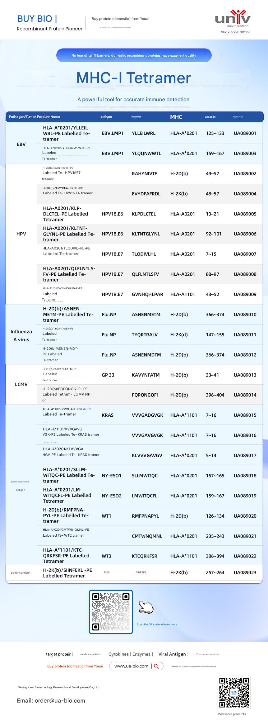
| Disease Category | Product Name | Antigen | Sequence | MHC | Position | Product Number |
|---|---|---|---|---|---|---|
| EBV | HLA-A*0201/YLELLVWRL-PE Labelled Tetramer | EBV.LMP1 | YLELLVWRL | HLA-A*0201 | 125-133 | UA089001 |
| EBV | HLA-A*0201/YLQQNWTL-PE Labelled Tetramer | EBV.LMP1 | YLQQNWTL | HLA-A*0201 | 159-167 | UA089003 |
| EBV | H-2Db(b)/RAHY-NIVTF-PE Labelled Tetramer | HPV16.E7 | RAHYNIVTF | H-2Db | 49-57 | UA089002 |
| HPV | H-2K(b)/EVYDFA-FRQL-PE Labelled Tetramer | HPV16.E6 | EVYDFARDL | H-2Kb | 48-57 | UA089004 |
| HPV | HLA-A*0201/KLP-DLCTL-PE Labelled Tetramer | HPV18.E6 | KLPDCTL | HLA-A*0201 | 13-21 | UA089005 |
| HPV | HLA-A*0201/KLTNT-GLYQL-PE Labelled Tetramer | HPV18.E6 | KLTNTGLYNL | HLA-A*0201 | 92-101 | UA089006 |
| HPV | HLA-A*0201/TLODIVIHL-PE Labelled Tetramer | HPV18.E7 | TLODIVIHL | HLA-A*0201 | 7~15 | UA089007 |
| HPV | HLA-A*0201/QFLNTL-FV-PE Labelled Tetramer | HPV18.E7 | QFLNTLFSV | HLA-A*0201 | 88-97 | UA089008 |
| HPV | HLA-A*1101/GVNHQLPAR-PE Labelled Tetramer | HPV18.E7 | GVNHQLPAR | HLA-A*1101 | 43-52 | UA089009 |
| Influenza A Virus | H-2D(b)/ASNENMETM-PE Labelled Tetramer | Flu.NP | ASNENMETM | H-2Db | 366-374 | UA089010 |
| Influenza A Virus | H-2K(d)/TYQR-TRALY-PE Labelled Tetramer | Flu.NP | TYQRTRALY | H-2Kd | 147-155 | UA089011 |
| Influenza A Virus | H-2D(b)/ASNEN-MDTM-PE Labelled Tetramer | Flu.NP | ASNENMDTM | H-2Db | 366-374 | UA089012 |
| LCMV | H-2D(b)/KAVYNFATM-PE Labelled Tetramer | GP 33 | KAVYNFATM | H-2Db | 33-41 | UA089013 |
| LCMV | H-2D(b)/FQPGQGFVK-PE Labelled Tetramer | LCMV NP | FQPGQGFVK | H-2Db | 396-404 | UA089014 |
| Tumor-related | HLA-A*1101/VVGADGVK-PE Labelled Tetramer | KRAS | VVGADGVK | HLA-A*1101 | 7~16 | UA089015 |
| Tumor-related | HLA-A*1101/VVGAGVGK-PE Labelled Tetramer | KRAS | VVGAGVGK | HLA-A*1101 | 7~16 | UA089016 |
| Tumor-related | HLA-A*0201/KLVVGAGV-PE Labelled Tetramer | KRAS | KLVVGAGV | HLA-A*0201 | 5~14 | UA089017 |
| Tumor-related | HLA-A*0201/SLLMWITQC-PE Labelled Tetramer | NY-ESO1 | SLLMWITQC | HLA-A*0201 | 157-165 | UA089018 |
| Melanoma | HLA-A*0201/LMWITQCFL-PE Labelled Tetramer | NY-ESO2 | LMWITQCFL | HLA-A*0201 | 159-167 | UA089019 |
| Melanoma | H-2Db(b)/MMFPNA-P1-PE Labelled Tetramer | WT1 | RMFPNAPL | H-2Db | 126-134 | UA089020 |
| Melanoma | HLA-A*0201/CMTWV-PE Labelled Tetramer | WT2 | CMTWVNMDM | HLA-A*0201 | 235-243 | UA089021 |
| Melanoma | HLA-A*1101/KTCQRKSF-PE Labelled Tetramer | WT3 | KTCQRKSF | HLA-A*1101 | 386-394 | UA089022 |
| Ovarian Cancer | H-2K(b)/SINFEKL-PE Labelled Tetramer | OVA | SINFEKL | H-2Kb | 257-264 | UA089023 |
This article is reviewed and published by the technical expert team of UA
Disclaimer: This article partially utilizes artificial intelligence assistance in its creation. If any content involves copyright or intellectual property issues, please let us know and we promise to verify and remove it as soon as possible.
Purchase recombinant protein, choose Nanjing UA-Bio
UA protein focuses on providing various protein reagents, raw materials, and services required for drug research and development, cell therapy, gene therapy, and basic scientific research, including drug target proteins, immune checkpoint proteins, cytokines, tool enzymes, customized protein expression, and full-length transmembrane protein development. Youai is committed to providing customers with high-quality products and professional services, and building a High-tech Biological Enterprise with International Competitiveness.







 Business license (three certificates in one)