I. Discovery and Biological Characteristics of LRRC15+ Fibroblasts
In the field of tumor research, the application of single-cell technology has promoted in-depth exploration of cellular heterogeneity in the tumor microenvironment. Recently, through single-cell analysis of mouse and human cancer samples, a subpopulation of fibroblasts highly expressing leucine-rich repeat-containing protein 15 (LRRC15) has been successfully identified. As an important component of the tumor microenvironment, this cell population has unique biological characteristics.
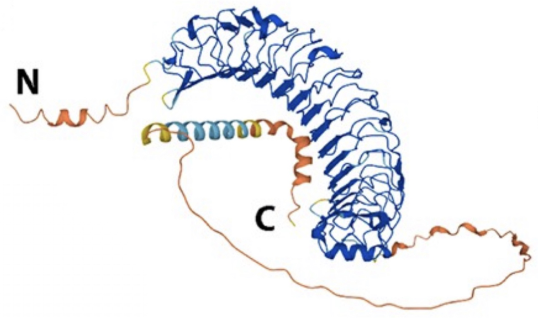
LRRC15 belongs to the leucine-rich repeat superfamily, and its molecular structure contains multiple leucine repeat motifs, which are usually involved in biological processes such as cell recognition, signal transduction, and protein interaction. In the discovered LRRC15+ fibroblasts, there is a large number of gene expressions related to extracellular matrix metabolism, accompanied by abnormal expressions of various immune regulatory molecules. These characteristics indicate that LRRC15 is not only a specific marker of this cell subpopulation but also may play a direct role in the regulation of cell functions. However, key issues such as the molecular function of LRRC15 itself, the regulatory mechanism of its specific expression in fibroblasts, and how it affects the dynamic balance of the tumor microenvironment remain to be further studied.
II. Differentiation Regulation Mechanism of LRRC15+ Fibroblasts
The differentiation and formation of LRRC15+ fibroblasts is a complex process regulated by multiple signaling pathways. Studies have shown that the transforming growth factor β (TGFβ) signaling pathway plays a core role in this process. By constructing a fibroblast-specific labeling model and combining gene editing technology to conditionally regulate the TGFβ receptor signaling pathway, experiments have confirmed that the differentiation of LRRC15+ fibroblasts is highly dependent on the activation of TGFβ signaling.
Gene expression profile analysis shows that in addition to highly expressing LRRC15, LRRC15+ fibroblasts also express classic cancer-associated fibroblast marker genes such as COL10A1, COL11A1, and MMP11, suggesting their biological properties as activated fibroblasts. Further studies have found that in fibroblasts with high LRRC15 expression, the expression levels of downstream target genes of the TGFβ signaling pathway are significantly upregulated, and the expression level of LRRC15 is positively correlated with TGFβ signaling activity. This result reveals that the TGFβ-LRRC15 regulatory axis may play a key role in the activation process of fibroblasts, providing important clues for understanding the biological origin of LRRC15+ fibroblasts.
III. Mechanism of Action of LRRC15+ Fibroblasts in Tumor Progression
(I) Promoting Effect on Tumor Growth
To clarify the function of LRRC15+ fibroblasts in tumor development, studies have adopted specific cell ablation technology to target and eliminate fibroblasts expressing LRRC15. Experimental results show that when LRRC15+ fibroblasts are effectively removed, the growth rate of tumor tissues slows down significantly, and the volume shrinks obviously. This finding directly proves that LRRC15+ fibroblasts have the effect of promoting tumor growth, suggesting that LRRC15 may become a potential biological marker for evaluating tumor progression.
In-depth analysis found that LRRC15+ fibroblasts can reshape the microenvironment around tumor cells by secreting various cytokines and growth factors, providing favorable conditions for tumor cell proliferation. At the same time, the highly expressed molecules such as matrix metalloproteinases can degrade the extracellular matrix and promote the invasive growth of tumor tissues. These research results provide experimental basis for the molecular mechanism by which LRRC15+ fibroblasts promote tumor growth.
(II) Regulatory Role in Tumor Immune Microenvironment
The role of LRRC15+ fibroblasts in tumor immune regulation has attracted increasing attention. Clinical studies have observed that tumor tissues enriched with LRRC15+ fibroblasts are often accompanied by the characteristics of immune desert-type microenvironment, showing a decrease in the infiltration of anti-tumor immune cells. Experimental studies through specific ablation models have further confirmed that LRRC15+ fibroblasts can affect anti-tumor immune responses by inhibiting the function of CD8+ T cells.
In the LRRC15+ fibroblast ablation model, the number and activity of CD8+ T cell infiltration in tumor tissues increased significantly, accompanied by inhibited tumor growth; while when CD8+ T cells were eliminated, the anti-tumor effect brought by LRRC15+ fibroblast ablation was completely reversed. This result reveals that LRRC15+ fibroblasts promote tumor immune escape by impairing CD8+ T cell-mediated adaptive immune responses. Further studies have found that LRRC15+ fibroblasts can secrete a variety of immunosuppressive factors and at the same time hinder immune cell infiltration by reshaping the extracellular matrix structure, jointly forming an immunosuppressive microenvironment.
(III) Impact on Tumor Immunotherapy Response
The difference in clinical response to immune checkpoint inhibitor therapy is an important challenge in tumor treatment, and LRRC15+ fibroblasts may be one of the key factors affecting the therapeutic effect. Animal experiments show that in tumor models with LRRC15+ fibroblasts, the effect of anti-PD-L1 antibody therapy is limited; while in models lacking LRRC15+ fibroblasts, the sensitivity of tumors to immunotherapy is significantly improved, and the treatment response rate is obviously increased.
The mechanism of this phenomenon may be related to the improvement of the immune microenvironment by LRRC15+ fibroblasts. After LRRC15+ fibroblasts are eliminated, the level of immunosuppressive cytokines in tumor tissues decreases, and the infiltration and activation status of CD8+ T cells are improved, enabling immune checkpoint inhibitors to more effectively activate anti-tumor immune responses. This suggests that the expression level of LRRC15 may become a potential biomarker for predicting tumor immunotherapy response, and targeting LRRC15+ fibroblasts may provide a new strategy for improving the efficacy of immunotherapy.
IV. Scientific Significance and Application Prospects of LRRC15 Research
As a specific marker of tumor-associated fibroblasts, LRRC15 has research value beyond simple cell typing. In-depth research on LRRC15+ fibroblasts not only reveals new mechanisms of cell-cell interactions in the tumor microenvironment but also provides a theoretical basis for the development of new tumor treatment strategies.
From the perspective of clinical transformation, LRRC15 may become a molecular marker for tumor diagnosis and prognosis evaluation, and its expression level can be used to guide the selection of individualized treatment plans. In terms of treatment strategies, targeted inhibition of the differentiation or function of LRRC15+ fibroblasts may play an anti-tumor role through dual mechanisms: directly inhibiting tumor growth and improving the tumor immune microenvironment to enhance the effect of immunotherapy. In the future, with the in-depth analysis of LRRC15 molecular functions and the development of specific targeted drugs, LRRC15-related treatment strategies are expected to provide new breakthroughs for precise tumor treatment.













 Business license (three certificates in one)