I. Molecular Characteristics and Biological Significance of MOG
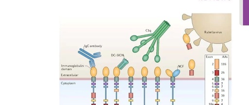
Myelin oligodendrocyte glycoprotein (MOG) is a glycoprotein specifically expressed on the surface of oligodendrocytes and the outermost layer of myelin in the central nervous system (CNS). As a member of the immunoglobulin superfamily, it consists of 218 amino acids. Despite its extremely low expression abundance in myelin components (< 0.05%), its physiological functions are crucial.
Based on structural characteristics, it is 推测 that MOG may be involved in maintaining myelin structural stability, regulating the dynamic balance of oligodendrocyte cytoskeleton, and activating the complement system and other physiological processes. More importantly, MOG is a key autoantigen. Animal experiments have confirmed that MOG antibody (MOG-Ab) is a pathogenic antibody in inflammatory demyelinating disease models, which can directly mediate myelin damage, providing a core target for the study of autoimmune demyelinating diseases.
II. Detection Technology of MOG-Ab and Definition of MOG-AD
(I) Evolution of Detection Methods
In the 1980s, researchers first discovered MOG-Ab in experimental autoimmune encephalomyelitis models and confirmed that it can exacerbate demyelinating lesions. Early detection used Escherichia coli-synthesized MOG extracellular domain as the antigen, detected by enzyme-linked immunosorbent assay or Western blotting, but only linear epitope antibodies could be identified. Such antibodies exist in both healthy individuals and multiple sclerosis (MS) patients and lack pathogenicity. Animal experiments have confirmed that only antibodies against conformational epitopes are pathogenic, and early methods have obvious missed detections.
The cell-based assay (CBA) significantly improves detection accuracy by transfecting MOG RNA to enable engineered cells to express correctly conformational MOG protein, followed by immunofluorescence detection of conformational epitope antibodies. In 2018, the international consensus formally recommended the CBA method as the standard detection method and clarified that the diagnosis of MOG-Ab associated disorders (MOG-AD) must be based on positive antibodies, combined with typical clinical manifestations and auxiliary examination results.
(II) Disease Definition of MOG-AD
With the advancement of detection technology, researchers have found that MOG-Ab positive patients have commonalities in clinical phenotype, disease course, imaging, and treatment response, and there are significant differences from MS and aquaporin 4 antibody (AQP4-Ab) positive neuromyelitis optica spectrum disorders (NMOSD). At the same time, immunological studies have confirmed the pathogenicity of human MOG-Ab, so the academic community generally recognizes that MOG-AD is an independent disease entity.
III. Pathogenesis and Pathological Features of MOG-AD
(I) Immunopathological Mechanism
MOG is only expressed in the immune-privileged CNS, and the mechanism by which it triggers peripheral immune responses has not been fully elucidated. It is currently 推测 that when the blood-brain barrier is damaged due to infection and other factors, MOG antigens may leak into the peripheral circulation and be recognized by the immune system. Animal studies have shown that intestinal flora can assist the activation of MOG-specific CD4⁺T cells and B cells, promoting the production of MOG-Ab.
When the blood-brain barrier is damaged again, peripheral MOG-Ab enters the CNS and attacks myelin structures through complement activation and antibody-mediated cytotoxicity, leading to demyelination. Local inflammatory responses at the lesion site recruit lymphocytes and macrophages to infiltrate, and the pro-inflammatory factors such as IL-12, IL-17, and tumor necrosis factor secreted by them further amplify the inflammatory cascade, exacerbating nerve damage.
(II) Pathological Tissue Features
Pathological studies of MOG-AD are mainly based on brain biopsy. Typical features include perivascular lymphocyte infiltration, myelin loss, accompanied by IgG and complement deposition and macrophage activation. A few cases show perivascular granuloma-like structures, and lesions involve brain parenchyma, leptomeninges and other parts. Although it has similarities with other inflammatory demyelinating diseases, its immune complex deposition pattern and cell infiltration characteristics are unique.
IV. Clinical Phenotypes and Imaging Features of MOG-AD
(I) Epidemiological Characteristics
MOG-AD has unique epidemiological characteristics: the positive rate of MOG-Ab in children with inflammatory demyelinating diseases is about 40%, significantly higher than 22% in adults; there are slightly more female patients than male patients (1:1.1), with no racial aggregation, in contrast to NMOSD. Clinical phenotypes are related to age. In children, acute disseminated encephalomyelitis (ADEM)-like manifestations are predominant, while in adults, optic neuritis (ON) is the most common. Other phenotypes include myelitis, brainstem encephalitis, meningitis, etc.
(II) Main Clinical Phenotypes
Optic Neuritis (ON): The most common phenotype, characterized by acute vision loss, accompanied by eye pain or pain on eye movement. More than half of the patients have bilateral optic nerve involvement at onset. MRI shows optic nerve thickening and swelling, hyperintensity on T2-weighted images, and enhanced scan shows optic nerve sheath enhancement, about half of which extends to orbital soft tissues, which is a specific imaging change. Most patients have good visual recovery after treatment, and the blindness rate is significantly lower than that of AQP4-Ab positive NMOSD.
Myelitis: About 20% of patients have spinal cord damage, manifested as limb paralysis, sensory and urinary and fecal dysfunction. Compared with NMOSD, lower thoracic segment and conus medullaris involvement are more common, and the incidence of erectile dysfunction is higher in male patients. 80% of patients have lesions longer than 3 vertebral segments, and non-continuous short-segment lesions may appear. MRI shows "H"-shaped hyperintensity on axial view and linear hyperintensity on the anterior side of the spinal cord on sagittal view, which has differential value.
Encephalitis and Meningitis: About 20.7% of patients present with encephalitis symptoms, including abnormal mental behavior, epilepsy and disturbance of consciousness; some patients with meningeal involvement have headache, fever and meningeal irritation signs, which are easily misdiagnosed as intracranial infection. Brain MRI is mainly ADEM-like changes, with more leptomeningeal enhancement, cortical and deep nuclear lesions. When involving infratentorial structures, dysarthria, ataxia, etc. appear.
V. Treatment Strategies and Prognosis of MOG-AD
The treatment of MOG-AD lacks evidence from large-scale clinical trials and is mainly based on small-sample studies and clinical experience. Methylprednisolone pulse therapy is preferred in the acute phase, followed by oral prednisone with slow tapering. For patients with poor response to 激素,combined use of intravenous immunoglobulin or plasma exchange can be considered. The disease has obvious steroid dependence, and most relapses occur during steroid tapering, which is closely related to the speed of tapering. When the tapering time is less than 3 months, the recurrence rate doubles. Therefore, it is recommended that the course of oral 激素 should be more than 3 months, followed by sequential immunosuppressant therapy. It should be noted that glatiramer acetate, beta interferon and natalizumab may aggravate the condition and are not recommended.
In general, the prognosis of MOG-AD is better than that of MS and AQP4-Ab positive NMOSD, with lower recurrence risk, better Expanded Disability Status Scale (EDSS) score and visual prognosis. However, 47% of patients still have permanent neurological deficits, and myelitis patients have a higher risk of disability than ON patients.
VI. Research Prospects
As a new type of autoimmune demyelinating disease, MOG-AD has made progress in research in recent years, but there are still many problems to be solved. In the future, it is necessary to further clarify the molecular mechanism of MOG-Ab production and breaking through the blood-brain barrier, improve multi-dimensional diagnostic criteria, and carry out large-scale randomized controlled trials to establish standardized treatment plans, promoting the development of precise diagnosis and treatment of MOG-AD.












 Business license (three certificates in one)