Nystagmus refers to involuntary movements of both eyes, manifested as shaking or jerking of the eyes. In canines, this condition also has various subtypes, including congenital nystagmus (also known as Infantile Nystagmus Syndrome, INS), latent or manifest nystagmus associated with strabismus (now called Fusional Maldevelopment Nystagmus Syndrome, FMNS), acquired nystagmus (Acquired Nystagmus, AN), and special types of nystagmus such as Spasmus Nutans. Although these nystagmus types all present as eye shaking under naked-eye observation, high-speed camera recordings at 1000 frames per second reveal significant differences in their movement patterns, with different nystagmus waveforms often corresponding to different etiologies.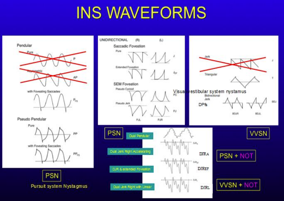
I. Overview of Canine Congenital Nystagmus
Canine congenital nystagmus refers to involuntary, rhythmic abnormal eye movements that occur in puppies within 6 months after birth. This abnormal eye movement can cause severe damage to a dog's visual function. Although there is a lack of comprehensive statistical data on the incidence of congenital nystagmus in the canine population, it is relatively common in veterinary clinical practice, especially in canine ophthalmology.
Currently, the etiology of canine congenital nystagmus has not been fully clarified. In recent years, genetic studies have found that mutations in some genes may be related to canine nystagmus, and abnormal retinal development is also considered a potential pathogenic factor. These studies provide certain clues for in-depth understanding of the pathogenesis of canine congenital nystagmus, but many issues remain to be further explored. In clinical practice, the understanding of canine nystagmus focuses more on its clinical manifestations and examination methods.
II. Eye Movement Recording Technology for Canine Nystagmus
On the basis of clinical examination of canine nystagmus, Eye Movement Recording (EMR) technology is a key examination method. Using devices such as EyeLink 1000 plus (with a sampling frequency of up to 1000Hz and an accuracy of 0.25 degrees), it can accurately capture the subtle characteristics of canine eye movements, providing rich information for diagnosis and evaluation.
Through EMR technology, the following important information about canine nystagmus can be obtained: first, the type of nystagmus, to clarify which subtype the dog's nystagmus belongs to; second, the amplitude of nystagmus, to understand the range of eye shaking; third, the frequency of nystagmus, that is, the number of eye shakes per unit time; fourth, the foveal fixation time of nystagmus, which is closely related to visual function; fifth, the null zone of nystagmus, that is, the fixation position with the smallest amplitude of nystagmus; sixth, the convergence damping of nystagmus; seventh, the periodic changes of nystagmus, such as Periodical Alternate Nystagmus (APAN); eighth, the nystagmus acuity function. Among them, nystagmus acuity function is an important new technology, which is of great significance for evaluating the visual function status of canine nystagmus.
III. Canine Nystagmus Acuity Function (NAF) Quantification Technology
Canine Nystagmus Acuity Function (NAF) quantitative analysis is calculated based on the speed, waveform, and fixation time of nystagmus movement, which can reflect the best visual acuity of dogs in the state of nystagmus. It is not a parameter that can be simply expressed by the frequency and amplitude of nystagmus. NAF quantitative analysis requires strict calibration of left and right eyes, and the equipment is required to have a sampling frequency of at least 250Hz.
At present, an accurate, fast, and easy-to-use Automated Nystagmus Acuity Function (ANAF) analysis software has been developed, which is suitable for application in veterinary clinics. ANAF analysis can sensitively reflect the subtle changes in canine nystagmus function. Whether conducting research on canine nystagmus or carrying out clinical diagnosis and treatment work, ANAF analysis technology is an indispensable important tool, providing reliable technical support for accurately evaluating the visual function status of canine nystagmus.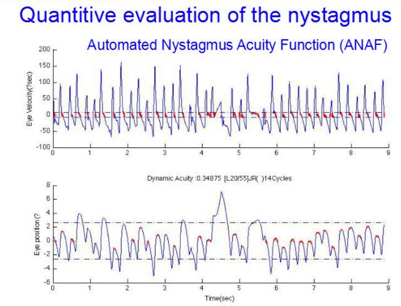
IV. New Advances in the Treatment of Canine Nystagmus
(I) Non-surgical Treatment Methods
In the non-surgical treatment of canine nystagmus, there are various exploration directions. Gene therapy has shown potential in improving visual function in some congenital eye diseases, providing new ideas for the treatment of canine congenital nystagmus; some drugs such as Baclofen, Gabapentin, and Memantine have shown certain efficacy in clinical application for individual types of canine congenital nystagmus; the topical ophthalmic drug Brinzolamide has been found to improve the nystagmus acuity function of dogs; prisms can also play a certain auxiliary role in the treatment of some affected dogs, helping to improve their visual experience; acupuncture, as a traditional therapy, can temporarily reduce nystagmus symptoms in dogs. However, for most canine nystagmus patients, surgical treatment is still needed to improve their condition.
(II) Surgical Treatment Methods
There are various surgical treatment methods for canine nystagmus, including the following common ones: The first is Null Zone Shifting, commonly known as the null zone transposition surgery, also known as compensatory head position correction surgery. By shortening and recession of the extraocular muscles, the dog's habitual fixation eye position (null zone) is rotated to the straight primary position, so as to improve its visual function and head posture. There are various types of compensatory head positions, including left or right face rotation, mandibular retraction or elevation, left or right head tilt, and even compound head positions.
The second surgical method is to create exophoria through surgery to induce convergence damping, thereby reducing the degree of nystagmus. The third is excessive recession of extraocular muscles, which can significantly reduce the intensity of nystagmus. The fourth is Tenotomy and Reattachment (T&R) of extraocular muscles, referred to as T&R surgery. This surgery does not change the position of the extraocular muscle insertion or the muscle strength, nor does it stimulate the adaptation mechanism of the eye movement system. It can improve nystagmus acuity function and reduce nystagmus intensity to a certain extent. It is believed that the therapeutic effect of this surgery is achieved by changing the input of extraocular muscle proprioceptors, but clinical practice shows that the therapeutic effect of this surgery is not satisfactory for all affected dogs. The fifth is injecting drugs into the extraocular muscles to enhance convergence damping or inject corresponding drugs into affected dogs at specific periods to reduce nystagmus.
V. Related Research Progress in Canine Nystagmus
In the field of canine nystagmus research, the relationship between proprioception and nystagmus is one of the hot topics, and numerous studies have conducted in-depth explorations on this. It is generally believed that the proprioceptive information of canine extraocular muscles comes from the Palisade endings of the extraocular muscles. This information is transmitted to the oculomotor control center through the trigeminal nerve, participating in the design and calculation of subsequent eye movements, and is the main afferent information source for oculomotor control. Facial sensory stimulation (including contact lenses, vibration, and electrical stimulation) shares the trigeminal nerve pathway with ocular proprioception, and reduces nystagmus symptoms by affecting the input of extraocular muscle proprioception.
However, recent studies have reported that contact lenses have no obvious effect on canine nystagmus, and there is also controversy about whether the function of the Palisade endings of extraocular muscles is sensory or motor, which challenges the theoretical basis of T&R surgery. Researchers have found that the number of Palisade endings in the extraocular muscles of dogs with nystagmus is increased, but it is not clear whether the proprioception of extraocular muscles is enhanced or weakened in the state of nystagmus. Currently, there is a lack of effective methods to detect ocular proprioception in clinical practice. If the correlation between canine nystagmus and proprioception can be clarified through research and a method to measure ocular proprioception can be established, it will help predict surgical prognosis according to the proprioceptive state of affected dogs and select the best treatment plan for them. It has been observed that tongue surface stimulation can cause a significant reduction in canine nystagmus, which is consistent with reports that facial sensory stimulation affects nystagmus. It is analyzed that this effect may be achieved through the participation of proprioception. However, why the impact varies among different affected dogs, with some nystagmus reduced by about 50% and others by only about 10%, and whether this is related to the functional state of proprioception, still needs further in-depth research to clarify the correlation between canine nystagmus and proprioception.












 Business license (three certificates in one)