




PDGF-AA: The "Mesenchymal Cell Regulatory Core" that drives tissue repair in PRP


PDGF-AA (a homodimer composed of two A subunits) has become a core functional molecule for PRP to promote wound healing and tissue regeneration due to its precise regulatory ability on mesenchymal cells (fibroblasts, mesenchymal stem cells, etc.). Its receptor binding properties and biological functions directly determine the repair efficiency of PRP
- Recent Advances
- Product Information
I. Introduction: The Role of PDGF-AA in the PRP Growth Factor Network
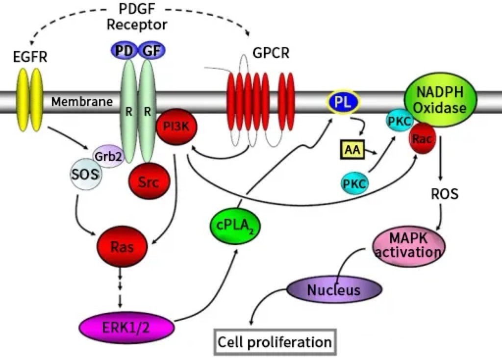
II. Molecular Characteristics and Expression Distribution of PDGF-AA
(I) Molecular Structure and Receptor Specificity
(II) Tissue and Cellular Expression Patterns
Embryonic stage: Secreted by epithelial cells (alveolar and gastrointestinal epithelium) and vascular smooth muscle precursor cells, participating in the development of the lungs and cardiovascular system;
Adult stage: Highly expressed in alveolar epithelium (lungs), placental trophoblasts, and prostatic glandular epithelium to maintain organ homeostasis; low-level secretion by dermal fibroblasts and bone marrow stromal cells supports tissue repair;
In PRP: Primarily stored in platelet α-granules, released upon activation by endogenous thrombin post-injection, serving as a core signaling source in the local repair microenvironment.
III. Core Biological Functions of PDGF-AA
(I) Regulating Mesenchymal Cell Proliferation and Chemotaxis
(II) Maintaining Organ Development and Homeostasis
Pulmonary function: Induces mesenchymal cell differentiation into alveolar smooth muscle cells during embryogenesis to maintain alveolar expansion; repairs alveolar damage in adulthood, slowing the pathological progression of chronic obstructive pulmonary disease (COPD);
Gastrointestinal homeostasis: Promotes collagen synthesis by intestinal mesenchymal fibroblasts to support epithelial cells and regulate the intestinal stem cell microenvironment;
Spermatogenesis: Stimulates the proliferation of testicular Leydig cells and testosterone synthesis, providing a hormonal environment for sperm maturation.
(III) Mediating Injury Repair and Regeneration
Inflammatory phase: Activates macrophages to phagocytose necrotic tissue and secrete inflammatory factors, amplifying repair signals;
Proliferative phase: Recruits fibroblasts to synthesize extracellular matrix (ECM), supports angiogenesis, and forms granulation tissue;
Remodeling phase: Induces fibroblast differentiation into myofibroblasts, regulates ECM remodeling, and reduces scarring. Preclinical studies show that PRP with high PDGF-AA concentration shortens mouse skin wound healing time by 30% and increases the tensile strength of healed skin by 25%.
IV. Abnormal PDGF-AA Expression and Associated Diseases
Tumors: In cholangiocarcinoma and head and neck squamous cell carcinoma (HNSCC), cancer cells overexpress PDGF-AA, promoting tumor stroma formation and vascular maturation, enhancing invasiveness, and reducing chemotherapy sensitivity;
Fibrosis: Post-lung injury, alveolar epithelial cells overproduce PDGF-AA, activating fibroblasts and leading to alveolar fibrosis; during renal ischemia, renal tubular epithelial cells secrete PDGF-AA, accelerating renal interstitial fibrosis;
Developmental defects: Mice with PDGF-A gene knockout (unable to synthesize PDGF-AA) exhibit defects in alveolar smooth muscle and testicular Leydig cells, mostly dying in the embryonic stage or within 60 days after birth.
V. Application and Outlook of PDGF-AA in PRP
Chronic wounds: Compensates for insufficient endogenous growth factors in patients with diabetic foot ulcers or pressure sores, accelerating healing;
Orthopedic repair: Promotes the proliferation of osteoprogenitor cells and migration of tendon cells, aiding bone transplantation and tendon repair;
Soft tissue regeneration: Drives skin collagen synthesis and oral mucosal renewal, improving repair quality.
VI. Conclusion
This article is reviewed and published by the technical expert team of UA
Disclaimer: This article partially utilizes artificial intelligence assistance in its creation. If any content involves copyright or intellectual property issues, please let us know and we promise to verify and remove it as soon as possible.
Purchase recombinant protein, choose Nanjing UA-Bio
UA protein focuses on providing various protein reagents, raw materials, and services required for drug research and development, cell therapy, gene therapy, and basic scientific research, including drug target proteins, immune checkpoint proteins, cytokines, tool enzymes, customized protein expression, and full-length transmembrane protein development. Youai is committed to providing customers with high-quality products and professional services, and building a High-tech Biological Enterprise with International Competitiveness.







 Business license (three certificates in one)