




HLA-A MHC Tetramer Technology: A Precision Key to Decoding Specific T-Cell Immune Responses


Prior to the 1990s, antigen-specific T cells were primarily detected using functional assays like LDA or ELISPOT, which indirectly measured T cell frequency through proliferation or cytokine secretion. These methods lacked TCR information, could not distinguish cell states, and had limited sensitivity for rare populations (<0.01%). Furthermore, their reliance on cell viability often altered native states, failing to accurately reflect the in vivo T cell repertoire.
- Recent Advances
- Product Information
I. What is MHC Tetramer Technology? What Scientific Challenges Did Its Emergence Address?
In the long history of immunological research, accurately identifying, quantifying, and isolating T cells specific to particular antigens (such as viral peptides or tumor neoantigens) has been a significant technical challenge. Before the 1990s, researchers primarily relied on functional assays, such as limiting dilution analysis (LDA) or enzyme-linked immunospot (ELISPOT), to indirectly estimate the number of specific T cells. These methods inferred the presence and function of T cells by observing their proliferative response or cytokine secretion (e.g., IFN-γ) upon antigen stimulation. However, these functional approaches had inherent limitations: they could not provide direct information about the T-cell receptor (TCR) on the T-cell surface, could not distinguish between memory and effector T-cell states, and had limited sensitivity, making it difficult to detect extremely low-frequency (<0.01%) antigen-specific T-cell populations. More importantly, functional assays depended on cell activity, and during the detection process, cells might undergo apoptosis, differentiation, or changes in functional state, thus failing to accurately reflect the initial T-cell repertoire in vivo.
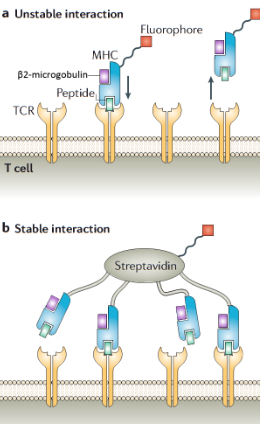
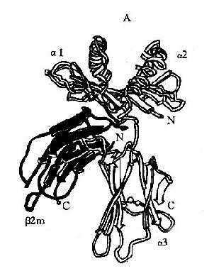
This dilemma was fundamentally resolved in 1996 by John Altman, Mark Davis, and others at the NIH. Inspired by the property of streptavidin to bind four biotin molecules with high affinity, they creatively applied this chemical system to immunological detection and invented MHC tetramer technology. The core principle is as follows: First, the heavy and light chains (β2-microglobulin) of the target MHC class I molecule (e.g., HLA-A*02:01) are produced using genetic engineering. Then, the extracellular domain of the heavy chain is fused with a specific substrate sequence for BirA enzyme (BirA substrate peptide) to enable site-specific biotinylation in vitro. Next, in the presence of a specific antigen peptide and β2-microglobulin, the biotinylated MHC heavy chain is correctly folded to form a stable pMHC complex. Finally, four identical pMHC monomer molecules are coupled with a fluorophore-labeled streptavidin via the biotin-streptavidin system, forming a fluorescently labeled, tetravalent pMHC complex—the MHC tetramer. This tetramer structure can bind with high affinity and specificity to T cells expressing the corresponding TCR on their surface. Its binding strength far exceeds that of monovalent or bivalent pMHC-TCR interactions, enabling the direct "visualization" and sorting of these specific T cells using flow cytometry. The birth of this technology provided immunologists with a high-precision "lens" to directly observe the true nature of T-cell immune responses, marking the beginning of a new era in T-cell immunological research with direct ex vivo detection.
II. What Is the Preparation Process of HLA-A MHC Tetramers? What Are the Key Technical Points?
Constructing an efficient, specific, and stable HLA-A MHC tetramer is a meticulous and multi-step biochemical process, where each step is critical and directly impacts the quality of the final product and the reliability of experimental results. The entire process begins with gene cloning and protein expression. Researchers must first obtain the coding sequence for the extracellular domain (α1, α2, α3 domains) of the target HLA-A allele (e.g., the widely prevalent HLA-A*02:01) and clone it into a prokaryotic (e.g., E. coli) or eukaryotic (e.g., insect cell-baculovirus system) expression vector. A key point is that a approximately 15-amino-acid BirA enzyme catalytic sequence must be fused to the C-terminus of the HLA heavy chain, which is a prerequisite for efficient and site-specific biotinylation. The gene for β2-microglobulin is typically cloned and expressed separately. The constructed vectors are then introduced into the expression system for large-scale production, followed by purification of soluble recombinant proteins through inclusion body refolding or directly from cell culture supernatants.
After obtaining purified HLA heavy chains, β2-microglobulin, and synthesized antigen peptides, the next step is the folding and purification of pMHC monomers. This is a highly delicate step that requires incubating the three components at precise molar ratios in an optimized redox buffer (often containing reagents like arginine to aid folding) to promote correct folding into stable pMHC complexes. Incorrectly folded protein aggregates and excess peptides are removed using techniques such as size-exclusion chromatography (SEC) or ion-exchange chromatography to obtain high-purity pMHC monomers. This is followed by the biotinylation reaction, where BirA enzyme is used to covalently attach biotin to a specific lysine residue within the BirA sequence at the C-terminus of the HLA heavy chain in the presence of ATP. The efficiency of the enzymatic reaction must be strictly monitored via electrophoresis or the HABA method to ensure near-100% biotinylation efficiency, as unbiotinylated monomers cannot form tetramers and become a source of background noise in subsequent detection.
The final step is tetramer assembly. Biotinylated pMHC monomers are mixed with fluorophore-labeled (most commonly PE, phycoerythrin, due to its extreme brightness) streptavidin at a molar ratio slightly higher than 4:1 and incubated on ice in the dark. The four biotin-binding sites of streptavidin "capture" four pMHC monomers, spontaneously assembling into a fluorescently labeled MHC tetramer. Excess monomers are removed through further gel filtration chromatography, resulting in a ready-to-use, highly homogeneous HLA-A MHC tetramer reagent that can be stored long-term at -80°C. The entire process demands extremely high standards for protein quality, folding efficiency, biotinylation efficiency, and assembly ratio control, representing a convergence of molecular biology, biochemistry, and immunology techniques.
III. What Revolutionary Applications Does This Technology Have in Basic and Clinical Research?
The advent of HLA-A MHC tetramer technology has significantly advanced nearly all related fields, from basic immunology to clinical immunotherapy. In basic research, it has become the "ultimate tool" for deciphering the dynamics of adaptive immune responses. Researchers can directly track the frequency, phenotype (e.g., memory subtypes: naive T cells, central memory T cells, effector memory T cells, terminally differentiated effector T cells), functional status (via combined intracellular cytokine staining), and TCR repertoire diversity of antigen-specific CD8+ T cell populations in peripheral blood, lymph nodes, or tumor-infiltrating lymphocytes (TILs) during viral infections (e.g., HIV, CMV, EBV, influenza), autoimmune diseases, or tumor development. This has made it possible to map the complete landscape of T-cell immune responses, such as revealing the kinetics of T-cell response expansion, contraction, and memory pool formation in acute infections, as well as the detailed characteristics of T-cell exhaustion in chronic infections and cancer.
In vaccine development, tetramer technology is the gold standard for assessing vaccine immunogenicity. Traditional antibody titer measurements only reflect the effects of humoral immunity, whereas the success of an effective vaccine, especially one aimed at preventing viral infections or treating cancer, hinges on its ability to induce a robust and lasting specific T-cell immune response. Using tetramer technology, researchers can precisely quantify the number and quality of antigen-specific CD8+ T cells produced in the body after vaccination, providing the most direct and reliable immunological basis for screening and optimizing vaccine candidates.
In tumor immunology and adoptive cell therapy (ACT), the role of tetramer technology is indispensable. It is widely used to identify and screen specific T-cell clones targeting tumor neoantigens or tumor-associated antigens (TAAs). Researchers can isolate these rare but potent anti-cancer T cells from patients, expand them in vitro, and reinfuse them into the patient—a approach known as TIL therapy or TCR-T cell therapy. Throughout the treatment process, tetramers are used to monitor the persistence, distribution, and functional status of the reinfused T cells in the patient, serving as a key biomarker for evaluating treatment efficacy and predicting prognosis. Additionally, in autoimmune disease research, tetramer technology has helped identify pathogenic T-cell clones that erroneously attack self-tissues, opening new avenues for understanding disease mechanisms and developing therapies to target and eliminate these pathogenic clones.
IV. Despite Its Power, What Major Challenges and Limitations Does MHC Tetramer Technology Currently Face?
Although MHC tetramer technology has brought revolutionary breakthroughs, it is not without its flaws, and its application still faces several important challenges and limitations. The primary challenge is its high HLA restriction. Tetramers are constructed based on specific HLA alleles (e.g., HLA-A*02:01) and can only detect T cells that recognize specific antigen peptides presented by that allele. Due to the extreme diversity of the HLA system, with significant genotypic differences across ethnicities and individuals, this means that tetramers must be individually designed and produced for different HLA types and antigen peptides, making the process costly and cumbersome and largely limiting its use for large-scale, unbiased screening applications.
Second, TCR affinity plays a role. Tetramer binding to T cells depends on the affinity of the pMHC-TCR interaction. For TCRs with very low affinity, even if they are biologically functional (capable of being activated by antigen), they may not form stable bonds, leading to false-negative results. Conversely, extremely high-affinity cross-reactivity may cause false positives. Additionally, the activation state of T cells can affect binding: some activated T cells may undergo TCR internalization or changes in surface molecule expression, impacting tetramer staining efficiency.
Third, background noise is an issue when detecting low-frequency T-cell populations. Especially when analyzing phenotypically complex samples (e.g., tumor tissue), non-specific binding or fluorescent signals from dead cells may mask the signals of truly antigen-specific T cells at very low frequencies, placing extremely high demands on instrument sensitivity and data analysis strategies.
Finally, traditional tetramer technology is a "targeted" technology; it can only be used to detect known T cells for known antigens and cannot discover or identify unknown new antigens or novel TCR specificities, meaning it has limited "non-hypothesis-driven" discovery capabilities. To overcome these limitations, a range of new technologies has emerged, such as high-throughput screening platforms based on DNA barcoding encoding multiple pMHCs, allowing simultaneous screening of T-cell responses to hundreds or even thousands of antigens in a single experiment, greatly expanding the application boundaries of tetramer technology.
V. What Are the Future Directions? How Will New Technologies Further Expand Its Potential?
To break through the bottlenecks of traditional tetramer technology, scientists are driving its iterative upgrade from multiple directions. A core direction is multiplexing and high-throughput. Metal-labeled tetramers based on mass cytometry (CyTOF) or multiplex pMHC tetramer combination technologies based on fluorescent barcoding enable the simultaneous detection of T-cell responses to dozens or even hundreds of different antigens in a single sample. This greatly improves detection efficiency, reduces sample requirements, and allows for the mapping of extremely detailed panoramic views of T-cell immune responses.
Another frontier is the integration of functional and dynamic detection. Combining tetramer staining with assays for cytokine secretion, proliferative capacity, killing activity, and transcriptomic and proteomic analyses goes beyond mere "counting" to deeply reveal the functional characteristics, differentiation trajectories, and metabolic states of these identified T cells, providing multi-dimensional information for assessing immune cell quality.
Furthermore, new structural designs are being explored. For example, using smaller fluorescent labels or novel fluorescent nanomaterials to reduce background; developing "cleavable" tetramers that can dissociate under specific conditions to address issues where high-affinity tetramers may artificially activate or interfere with T-cell function; and designing MHC class II tetramers to better study CD4+ T-cell responses.
Looking ahead, HLA-A MHC tetramer technology, as a milestone in immunological research, will continue to serve as the cornerstone for deciphering T-cell immunity. Its integration with cutting-edge technologies such as single-cell sequencing, CRISPR screening, and bioinformatics will undoubtedly drive deeper understanding and breakthrough clinical advances in fields such as infection immunity, tumor immunology, autoimmune diseases, and vaccine design, ultimately laying a solid technical foundation for precision immune monitoring and personalized immunotherapy.
Click on the product catalog numbers below to access detailed information on our official website.
This article is reviewed and published by the technical expert team of UA
Disclaimer: This article partially utilizes artificial intelligence assistance in its creation. If any content involves copyright or intellectual property issues, please let us know and we promise to verify and remove it as soon as possible.
Purchase recombinant protein, choose Nanjing UA-Bio
UA protein focuses on providing various protein reagents, raw materials, and services required for drug research and development, cell therapy, gene therapy, and basic scientific research, including drug target proteins, immune checkpoint proteins, cytokines, tool enzymes, customized protein expression, and full-length transmembrane protein development. Youai is committed to providing customers with high-quality products and professional services, and building a High-tech Biological Enterprise with International Competitiveness.







 Business license (three certificates in one)