Immune checkpoints are core molecules that maintain the homeostasis of immune responses, divided into "adaptive immune checkpoints" (regulating T cells and other cells) and "innate immune checkpoints" (regulating macrophages and other cells). As a key innate immune checkpoint, signal regulatory protein α (SIRP-α/CD172A) forms a "don’t eat me" signaling pathway by binding to its ligand CD47. Under physiological conditions, this pathway protects healthy cells from excessive phagocytosis; however, tumor cells hijack this pathway to evade immune clearance. In recent years, blocking strategies targeting the SIRP-α/CD47 axis have provided a new direction for tumors unresponsive to traditional treatments. This article focuses on its molecular characteristics, tumor evasion mechanisms, and research progress in blocking therapies.
SIRP-α is a type I transmembrane glycoprotein, with its core structure consisting of three parts: the extracellular region contains 1 IgV domain (the key region for binding to CD47) and 2 IgC2 domains; the transmembrane region has positively charged arginine residues, which participate in the initial regulation of signals; the intracellular region contains 2 ITIM motifs, the "core switch" for inhibitory functions. After binding to CD47, the tyrosine residues in ITIM are phosphorylated, recruiting SHP-1/2 phosphatases to inhibit cell activation. Its encoding gene PTPNS1 is located at 20p13 and has multiple splice variants, which regulate the tissue-specificity of inhibitory activity.
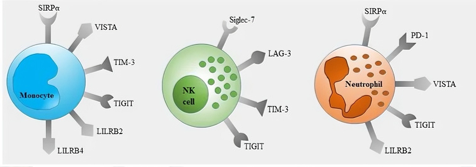
SIRP-α exhibits strong cell-type specificity in expression: it is mainly highly expressed in monocytes, macrophages (including tumor-associated macrophages, TAMs), neutrophils, and myeloid dendritic cells, directly associated with phagocytic function; it is also expressed in neuronal cells, presumably involved in neuroimmune homeostasis, though the mechanism remains unclear; in contrast, it is poorly expressed or absent in adaptive immune cells such as T cells and B cells, making it a unique innate immune checkpoint distinct from targets like PD-1.
The ligand CD47 is widely distributed on the surface of human cells, while tumor cells (e.g., acute myeloid leukemia, lung cancer, colorectal cancer) often abnormally overexpress CD47, forming an excessive "don’t eat me" signal and providing a specific basis for targeted therapy.
Under normal conditions, the SIRP-α/CD47 axis acts as a "brake system" for macrophages: CD47 on healthy cells binds to SIRP-α on macrophages, activating the ITIM-SHP-1/2 signal and inhibiting phagocytosis-related pathways such as PI3K-AKT. This prevents erroneous phagocytosis of red blood cells and autologous tissue cells, maintaining tissue integrity—for example, protecting red blood cells from excessive clearance by splenic macrophages.
Tumor cells achieve immune evasion by enhancing the SIRP-α/CD47 axis: first, they overexpress CD47. Clinical studies have shown that CD47 expression in tumor tissues is much higher than in normal tissues, and high expression is associated with poor patient prognosis (e.g., CD47 expression on AML cells is 3–5 times that of normal hematopoietic stem cells); second, the "don’t eat me" signal dominates phagocytic regulation. CD47 on tumor cells binds efficiently to SIRP-α on M2-type TAMs, continuously inhibiting phagocytosis. At the same time, tumors secrete cytokines such as IL-10 to induce macrophage polarization and upregulate SIRP-α, forming an "inhibitory loop"; third, they indirectly weaken adaptive immunity. Suppressed macrophages have reduced antigen-presenting ability and cannot activate CD8⁺ T cells, leading to dual immune evasion.
These antibodies bind to the IgV domain of SIRP-α, blocking its binding to CD47 or inducing SIRP-α degradation. In preclinical studies, a humanized anti-SIRP-α antibody increased macrophage phagocytic activity by 40%–60% in a mouse colorectal cancer model. When combined with anti-PD-1 antibodies, the tumor regression rate doubled compared to the single-drug group, with no significant hematological toxicity. Its advantages lie in strong targeting and high safety.
These antibodies bind to CD47 on tumor cells, preventing its binding to SIRP-α. Some antibodies also recruit macrophages to phagocytose tumors through antibody-dependent cellular phagocytosis (ADCP). Multiple drugs have entered Phase I/II clinical trials. For example, a candidate drug achieved an objective response rate (ORR) of 35% as monotherapy in relapsed/refractory AML, and the ORR increased to 58% when combined with azacitidine. However, it should be noted that CD47 on red blood cells may be bound, causing transient anemia, so antibody optimization is needed to reduce toxicity.
Bispecific molecules such as "anti-SIRP-α-anti-CD20" bispecific antibodies can target CD20 on B-cell lymphoma and SIRP-α on macrophages, enhancing phagocytosis; "anti-CD47-anti-PD-L1" bispecific antibodies synergistically activate macrophages and CD8⁺ T cells, achieving a tumor inhibition rate of over 80% in a mouse lung cancer model. SIRP-α-Fc fusion proteins enhance binding to CD47 through the Fc segment, competitively blocking the pathway. Without effector functions, they reduce damage to normal cells and shrink tumors by 65%–70% in solid tumor models when combined with chemotherapy.
Among tumor-intrinsic factors, patients with high CD47 expression are more sensitive, tumors with a high proportion of M1-type macrophages have better efficacy, and hematological tumors respond faster than solid tumors; among host factors, elderly patients or those with chronic inflammation have low myeloid cell activity, and intestinal flora imbalance can upregulate SIRP-α on macrophages, reducing drug efficacy.
SIRP-α/CD172A is a key "innate checkpoint" in tumor immune evasion, and related blocking strategies have shown anti-tumor potential in preclinical and early clinical studies. In the future, it is necessary to optimize drugs to reduce toxicity, screen biomarkers, and develop combination regimens, promoting this target to become an important supplement to tumor immunotherapy and bringing hope to more patients.












 Business license (three certificates in one)