




The potential of using tumor associated MHC tetramers to unlock new antigen research


Tumor associated antigens (TAAs) are commonly used for diagnosis, but they also exist in normal tissues, limiting their therapeutic value. In contrast, neoantigens - peptides derived from tumor specific gene mutations - are only expressed in cancer cells. This unique property makes it an ideal target for eradicating tumors with minimal side effects.
- Recent Advances
- Product Information
Introduction
Over the past decade, cancer immunotherapy has emerged as a breakthrough treatment alongside surgery, chemotherapy, and radiotherapy. By harnessing the immune system to target and eliminate cancer cells, immunotherapies have demonstrated remarkable efficacy across multiple cancer types. While tumor-associated antigens (TAAs) are commonly used in diagnostics, they are also present in normal tissues, limiting their therapeutic utility. In contrast, neoantigens—mutant peptides derived from tumor-specific genetic alterations—are expressed exclusively in cancer cells. This unique property makes them ideal targets for therapies aimed at eradicating tumors with minimal side effects. But how can we effectively identify and target these neoantigens? One powerful tool is the MHC tetramer, a technology that enables precise detection and analysis of neoantigen-specific T cells.
What Are Neoantigens and How Are They Presented?
Neoantigens are foreign proteins absent in normal tissues but produced in tumors through mechanisms such as genomic mutations, aberrant RNA splicing, post-translational modifications, or viral open reading frames. These antigens are processed and presented by major histocompatibility complex (MHC) molecules on tumor cells.
The journey of a neoantigen begins when it is digested into peptides by the proteasome. These peptides are transported into the endoplasmic reticulum (ER) via TAP (Transporters associated with antigen processing) proteins. In the ER, they bind to MHC-I molecules. For MHC-II presentation, the peptides are loaded onto MHC-II complexes in endosomal compartments after degradation of the invariant chain. The resulting peptide-MHC (pMHC) complexes are then displayed on the cell surface, where they can be recognized by T cells via their T cell receptors (TCRs).
This intricate process highlights the importance of MHC tetramers—multimeric pMHC complexes—in visualizing and isolating T cells that recognize specific neoantigens. By fluorescently labeling these tetramers, researchers can identify and study rare neoantigen-specific T cells with high precision.
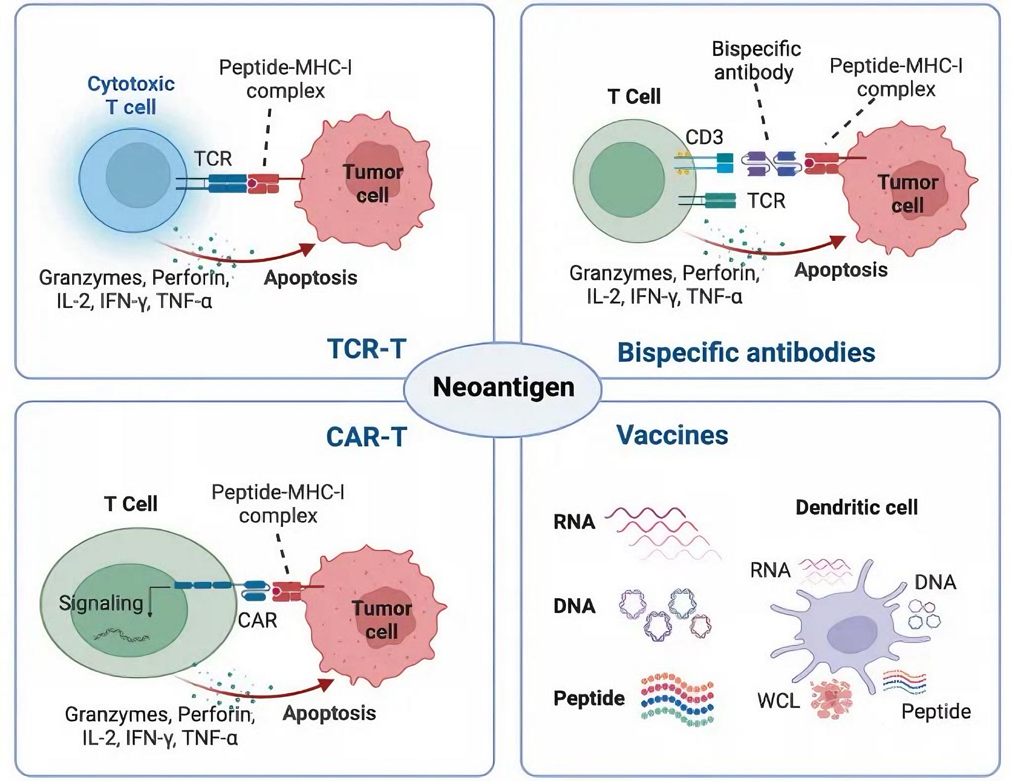
Why Are Neoantigens Ideal Targets for Immunotherapy?
Neoantigens offer two key advantages: tumor specificity and immunogenicity. Since they are not expressed in healthy tissues, targeting neoantigens minimizes off-target toxicity. Additionally, T cells targeting neoantigens escape central tolerance mechanisms, making them potent mediators of anti-tumor immunity.
Neoantigens can be classified into two types:
Private neoantigens: Unique to individual patients.
Public neoantigens: Shared across multiple patients and cancer types.
While private neoantigens enable personalized therapies, public neoantigens offer opportunities for "off-the-shelf" treatments. However, their identification requires sophisticated tools like MHC tetramers to validate T cell responses and guide therapeutic development.
How Are Immunogenic Neoantigens Identified and Validated?
The first step in leveraging neoantigens is identifying immunogenic candidates. Advances in next-generation sequencing (NGS), including whole-exome sequencing (WES) and RNA-seq, have enabled comprehensive profiling of tumor-specific mutations. However, not all mutations generate immunogenic neoantigens. Critical factors include:
Peptide processing and MHC presentation.
Affinity between pMHC complexes and TCRs.
Computational tools like NetMHC and NetCTLpan are used to predict MHC binding affinity, while mass spectrometry (MS) directly identifies MHC-bound peptides. Yet, computational predictions alone are insufficient. Experimental validation using MHC tetramers, ELISpot assays, or T cell activation assays is essential to confirm immunogenicity.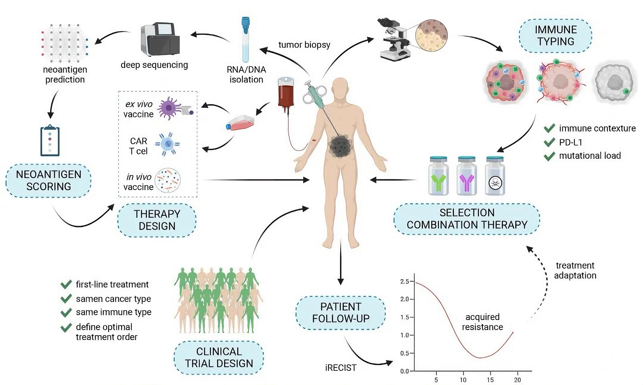
How Can MHC Tetramers Advance Neoantigen Research?
MHC tetramers are invaluable for:
Detecting neoantigen-specific T cells: By staining T cells with fluorescently labeled tetramers, researchers can quantify and isolate rare populations.
Evaluating T cell function: Combining tetramer staining with functional assays (e.g., cytokine production) provides insights into T cell efficacy.
Monitoring immunotherapy responses: Tetramers can track dynamic changes in neoantigen-specific T cells during treatment.
For example, in adoptive cell therapy (ACT), tetramers help identify and expand reactive T cells. In vaccine development, they assess immune responses to neoantigen-based vaccines.
What Are the Challenges and Future Directions?
Despite their promise, several challenges remain:
Tumor heterogeneity: Neoantigen expression varies within tumors, necessitating multiplexed tetramer approaches.
TCR affinity: Low-affinity interactions may be missed by standard tetramers.
Technical complexity: Tetramer production requires specialized expertise and resources.
Future innovations may include:
High-throughput tetramer screening to profile diverse neoantigen libraries.
Integration with single-cell technologies to link T cell specificity with functional states.
Development of personalized tetramers for clinical monitoring.
Conclusion
Neoantigens represent a frontier in cancer immunotherapy, offering unparalleled specificity and efficacy. The use of MHC tetramers is pivotal in translating neoantigen discoveries into therapies, from validating immunogenicity to guiding patient stratification. As technologies evolve, combining tetramers with multi-omics approaches will deepen our understanding of anti-tumor immunity and accelerate the development of next-generation immunotherapies.
This article is reviewed and published by the technical expert team of UA
Disclaimer: This article partially utilizes artificial intelligence assistance in its creation. If any content involves copyright or intellectual property issues, please let us know and we promise to verify and remove it as soon as possible.
Purchase recombinant protein, choose Nanjing UA-Bio
UA protein focuses on providing various protein reagents, raw materials, and services required for drug research and development, cell therapy, gene therapy, and basic scientific research, including drug target proteins, immune checkpoint proteins, cytokines, tool enzymes, customized protein expression, and full-length transmembrane protein development. Youai is committed to providing customers with high-quality products and professional services, and building a High-tech Biological Enterprise with International Competitiveness.







 Business license (three certificates in one)