




Potential Therapeutic Targets in Tumor Treatment - Siglec Family Proteins


Siglec, an acronym for Sialic acid-binding immunoglobulin-like lectins, represents a family of receptors that recognize sialylated glycans. The Siglec protein family is predominantly expressed on the surface of immune cells and is characterized by an immunoglobulin-like domain capable of binding sialic acids. These proteins play critical roles in mediating cell-cell interactions, as well as host-pathogen and host-tissue interactions, during processes such as infection and immune regulation.
- Recent Advances
- Product Information
- Reference
Siglec, an acronym for Sialic acid-binding immunoglobulin-like lectins, represents a family of receptors that recognize sialylated glycans. The Siglec protein family is predominantly expressed on the surface of immune cells and is characterized by an immunoglobulin-like domain capable of binding sialic acids. These proteins play critical roles in mediating cell-cell interactions, as well as host-pathogen and host-tissue interactions, during processes such as infection and immune regulation.
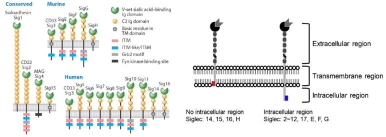
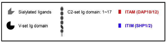
Figure 1: Schematic Diagram of Siglec Protein Structure
The Siglec family proteins feature an N-terminal immunoglobulin (Ig) domain, with the "variable region" of this domain containing the sialic acid-specific binding site. Additionally, they include repetitive "constant" C-regions of varying lengths. The transmembrane region contains amino acids that interact with motifs in the cell membrane. The intracellular region typically contains an immunoreceptor tyrosine-based inhibitory motif (ITIM). Most Siglec molecules possess these three domains. However, studies have shown that some Siglec molecules lack intracellular domains but instead contain immunoreceptor tyrosine-based activation motifs (ITAMs) in their transmembrane regions, such as Siglec-H, -14, -15, and -16.

Figure 2: Siglec-Expressing Cells and Associated Diseases
The Siglec family encompasses three primary functions:
Pathogen Evasion: Siglecs may be hijacked by sialic acid-bearing pathogens, facilitating infection or immune evasion.
Immune Cell Interaction and Function: For example, Siglec-1 (CD169) mediates antigen presentation by macrophages to T cells, Siglec-2 (CD22) regulates B cell differentiation, Siglec-3 (CD33) is involved in myeloid progenitor differentiation, and Siglec-7 and Siglec-9 modulate neutrophil apoptosis and phagocytosis.
ITIM/ITIM-like Motif Phosphorylation: Phosphorylated motifs recruit tyrosine phosphatases, thereby inhibiting immune activation.
Based on sequence homology, Siglecs are divided into two families:
Evolutionarily conserved classical Siglecs, including Siglec-1, Siglec-2 (CD22), Siglec-4, and Siglec-15, which possess the three aforementioned domains.
CD33-related Siglecs, which lack conserved regions. Human CD33-related Siglecs (Siglec-3, -5, -6, -7, -8, -9, -10, -11, -14, -16, -17) and murine CD33-related Siglecs (Siglec-3, -E, -F, -G, -H) exhibit 50%-90% sequence homology but differ in species-specific expression.
Siglec-2 (CD22) is highly expressed on B cells and is evolutionarily conserved in mammals. As an immune inhibitory receptor, it contains multiple intracellular ITIM motifs. By interacting with the B cell receptor (BCR), CD22 suppresses B cell responses and downregulates B cell activation, protecting the body from hyperactive B cells and excessive immune responses. This function is independent of CD22's sialic acid-binding ability. However, CD22's sialic acid-binding activity can attenuate its inhibitory effects. CD22 exhibits high specificity for α2-6-linked sialylated glycans. Mutations in the variable region arginine residue abolish sialic acid binding, leading to enhanced suppression of BCR function.
Siglec-3 (CD33) is an immune inhibitory receptor expressed on myeloid progenitor cells, monocytes, macrophages, dendritic cells, mast cells, and brain microglia. CD33 is highly expressed in acute myeloid leukemia (AML), and cytotoxic CD33 antibodies have been developed for cancer therapy. CD33 primarily binds α2-3- and α2-6-linked sialic acids.
Siglec-6 is predominantly expressed on placental trophoblasts during normal and abnormal embryonic pregnancies, as well as on peripheral leukocytes and intestinal tissues. Antibodies targeting Siglec-6 have been patented for inhibiting C3a-induced mast cell activation. Siglec-6 expression has been observed in AML cell lines, primary AML samples, and chronic B cell transformations.
Siglec-8 is primarily expressed on eosinophils and mast cells. Antibodies targeting Siglec-F (the murine homolog of human Siglec-8) induce apoptosis and eliminate eosinophils in mice. In patients with systemic mastocytosis, anti-Siglec-8 antibodies induce antibody-dependent cellular cytotoxicity against mast cells. These antibodies are also proposed for treating allergic diseases, including bronchial asthma. Autoantibodies against Siglec in intravenous immunoglobulin preparations have been reported to mediate anti-autoimmune and anti-allergic effects.
Siglec-9 is an immune inhibitory protein widely expressed on human leukocytes, monocytes, macrophages, neutrophils, dendritic cells, NK cell subsets, B cells, and T cells. Monoclonal antibodies or multivalent sialylated glycans that cluster Siglec-9 ligands on neutrophils induce neutrophil death, which is enhanced upon neutrophil activation. Siglec-9 binding to macrophages inhibits phagocytosis. Siglec-9 exhibits broad specificity for synthetic sialic acids in the presence of α2-3- and α2-6-linked sialylated glycans.
Siglec-15 has emerged as a significant target in cancer therapy. Its transmembrane region contains a positively charged amino acid residue, classifying it as an activating Siglec. Siglec-15 is expressed on tumor-associated macrophages (TAMs) in the tumor microenvironment (TME) of human lung, colorectal, and hepatocellular carcinomas. Interactions between Siglec-15-expressing THP-1 monocytes and sialic acid-expressing H157 lung cancer cells increase TGF-β production.
Siglec-15 is also expressed on stromal and cancer cells, including those in lymphoma, gastric cancer, and AML. Studies by Chen et al. demonstrate that Siglec-15 glycosylation affects its localization. Treatment with various lysosomal inhibitors reveals that Siglec-15 undergoes lysosome-dependent degradation in overexpressing HEK293 cells. Additionally, tunicamycin-mediated inhibition of glycosylation reduces Siglec-15 trafficking to the cell membrane and promotes lysosomal degradation.
UA BIOSCIENCE Siglec-Related Products
Click on the product catalog numbers below to be redirected to the official website for more details.
This article is reviewed and published by the technical expert team of UA
Disclaimer: This article partially utilizes artificial intelligence assistance in its creation. If any content involves copyright or intellectual property issues, please let us know and we promise to verify and remove it as soon as possible.
Purchase recombinant protein, choose Nanjing UA-Bio
UA protein focuses on providing various protein reagents, raw materials, and services required for drug research and development, cell therapy, gene therapy, and basic scientific research, including drug target proteins, immune checkpoint proteins, cytokines, tool enzymes, customized protein expression, and full-length transmembrane protein development. Youai is committed to providing customers with high-quality products and professional services, and building a High-tech Biological Enterprise with International Competitiveness.

1.Siglec-Ligand. Anabel Gonzalez-Gil.2021.
2.Functions and therapeutic targets of Siglec-mediated infections, inflammations and cancers. Chia-Hsueh Lin.2021.
3.Siglec Signaling in the Tumor Microenvironment. Eline J. H. van Houtum.2021.






 Business license (three certificates in one)