




Can platelet lysate replace fetal bovine serum? In depth analysis of the role and controversy of PDGF-AA


PDGF-AA (platelet-derived growth factor AA) is one of the key growth factors stored in platelet alpha granules and belongs to the PDGF family. In HPL, PDGF-AA plays a central role in regulating tissue repair and regeneration by promoting cell proliferation, migration, and synthesis of extracellular matrix.
- Recent Advances
- Product Information
1. What is platelet lysate, and how does it fundamentally differ from fetal bovine serum?
Human Platelet Lysate (HPL) is derived from human platelets and prepared by physically or chemically stimulating platelets (through repeated freeze-thaw cycles, ultrasonic treatment, or the addition of calcium ions/thrombin) to degranulate and release stored growth factors and cytokines, forming a liquid supplement. In contrast, Fetal Bovine Serum (FBS) is extracted from the blood of bovine fetuses and has long been widely used as a growth supplement in cell culture.
The most fundamental differences lie in their origin and composition: HPL is entirely human-derived, making its components more aligned with the physiological environment of human cells. It is particularly rich in various growth factors such as PDGF-AA, VEGF, and TGF-β. On the other hand, FBS is a xenogeneic serum. While nutrient-rich, it carries risks of introducing animal-derived pathogens, triggering immune reactions, and exhibiting significant batch-to-batch variability. Especially in the fields of cell therapy and tissue engineering, using animal-component-free HPL is regarded as a key strategy to enhance clinical safety.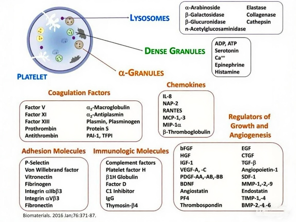
2. What role does PDGF-AA play in platelet lysate?
PDGF-AA (Platelet-Derived Growth Factor-AA) is one of the key growth factors stored in the α-granules of platelets and belongs to the PDGF family. In HPL, PDGF-AA plays a central role in regulating tissue repair and regeneration by promoting cell proliferation, migration, and the synthesis of extracellular matrix. For example, it significantly enhances the expansion capacity of mesenchymal stem cells (MSCs) and is involved in activating multiple signaling pathways related to angiogenesis and wound healing.
Studies have shown that after binding to its receptor PDGFR-α, PDGF-AA initiates downstream signaling pathways such as RAS-MAPK and PI3K-AKT, directly influencing cell cycle progression. This also explains why culture media supplemented with HPL often support more efficient MSC proliferation than traditional FBS-based cultures.
3. Can HPL fully replace FBS in cell culture applications?
Multiple studies have demonstrated that HPL performs excellently in supporting the in vitro expansion of various human cells (especially MSCs), even showing significantly higher proliferation efficiency than FBS. For instance, in some experiments, adipose-derived MSCs cultured in 20% HPL exhibited optimal proliferation while maintaining genomic stability and multidifferentiation potential.
However, HPL is not a "perfect substitute." Its biggest issue lies in considerable batch-to-batch variability. Due to differences among donors, platelet preparation methods, and activation techniques, different batches of HPL can vary significantly in the concentration and composition of growth factors. This inconsistency may sometimes lead to changes in cell behavior (such as immunomodulatory function or osteogenic/adipogenic differentiation capacity). Therefore, although HPL has clear advantages in avoiding animal-derived components, key challenges in standardization and quality control must be addressed before it can fully replace FBS.
4. How can high-quality platelet lysate be prepared? What are the pros and cons of different methods?
Various methods are used to prepare HPL, common strategies include repeated freeze-thaw cycles, thrombin activation, ultrasonic disruption, and solvent/detergent viral inactivation treatments. The freeze-thaw method is widely adopted due to its simplicity and low cost, but it may somewhat affect the activity of growth factors. Ultrasonic treatment is efficient but requires precise control to avoid protein denaturation. Chemical activation methods (such as adding CaCl₂ or thrombin) rapidly release factors but may introduce exogenous components, complicating subsequent purification and regulatory approval.
It is important to note that different preparation techniques directly impact the content and stability of key factors like PDGF-AA, thereby influencing the effectiveness of HPL in promoting cell proliferation. Therefore, when selecting an HPL preparation method, it is essential to consider the target cell type, application scenario, and compliance requirements.
5. Is it more appropriate to use autologous or allogeneic HPL?
HPL can be categorized into autologous (from the patient themselves) and allogeneic (from blood donor pools) based on its source. The greatest advantage of autologous HPL is that it eliminates the risk of infectious disease transmission and offers excellent immunocompatibility. However, its preparation is limited by the patient’s own platelet count and health status, making large-scale production challenging.
Allogeneic HPL, usually derived from pooled samples of multiple donors, is more conducive to mass production and a certain degree of standardization. Pathogen testing and inactivation treatments (such as S/D treatment) can reduce the risk of virus transmission. However, its potential immunogenicity and inter-donor variability cannot be overlooked. Particularly, ABO antigens present in the plasma of donors with different blood types may affect the immune characteristics of cultured cells if not handled properly.
6. How do HPL, PRP, and PRF differ? Do they all contain PDGF-AA?
Although Platelet-Rich Plasma (PRP), Platelet-Rich Fibrin (PRF), and HPL all originate from platelets, they differ significantly in preparation methods, composition, and application. PRP is a platelet concentrate obtained through centrifugation and still requires activation to release factors. PRF is formed through natural coagulation, creating a fibrin network containing platelets and leukocytes. HPL is the end product after platelets have been lysed and factors released, thus typically having a higher concentration of growth factors.
All three contain growth factors such as PDGF-AA, but HPL—with its factors already released into the plasma—is more straightforward to use directly in cell culture media and has a more homogeneous composition. It is important to note that PRP and PRF themselves do not contain stem cells and should not be misrepresented as "stem cell therapies."
7. Summary: Prospects and Challenges in the Application of HPL
Overall, due to its human-derived components, rich growth factors (such as PDGF-AA), and strong ability to support cell proliferation, Human Platelet Lysate (HPL) is a highly promising alternative to Fetal Bovine Serum. This is particularly relevant for meeting the demand for "animal-component-free" culture systems as cell therapy products move toward clinical translation.
However, challenges such as batch consistency, pathogen safety, impacts on various cellular functional properties, and the achievement of large-scale standardized production remain urgent issues to address. With future optimizations in preparation processes, strengthened quality control, and more in-depth research on the mechanisms of key factors like PDGF-AA, HPL is poised to play an increasingly important role in regenerative medicine and cell manufacturing.
This article is reviewed and published by the technical expert team of UA
Disclaimer: This article partially utilizes artificial intelligence assistance in its creation. If any content involves copyright or intellectual property issues, please let us know and we promise to verify and remove it as soon as possible.
Purchase recombinant protein, choose Nanjing UA-Bio
UA protein focuses on providing various protein reagents, raw materials, and services required for drug research and development, cell therapy, gene therapy, and basic scientific research, including drug target proteins, immune checkpoint proteins, cytokines, tool enzymes, customized protein expression, and full-length transmembrane protein development. Youai is committed to providing customers with high-quality products and professional services, and building a High-tech Biological Enterprise with International Competitiveness.







 Business license (three certificates in one)