




Navigating RAW264.7 macrophage polarization? Partner with U&I Bio to clearly define M1/M2.


Macrophage polarization refers to the process by which macrophages, stimulated by specific signals in the microenvironment, undergo functional reprogramming and differentiate into distinct subsets with different phenotypes and functions. This is not a simple "on" or "off" switch, but rather a complex functional spectrum, with the most classic types being M1 (classically activated) and M2 (alternatively activated).
- Recent Advances
- Product Information
In the world of immunological research, macrophages are undoubtedly the "versatile players" on the battlefield, capable of both charging into battle to clear pathogens and calming the aftermath to repair tissue. The mouse RAW264.7 cell line, serving as a "star model" for in vitro studies of macrophage function, requires precise regulation of its polarization, which is key to uncovering the core mechanisms of life processes such as inflammation, infection, cancer, and tissue repair.
Part 1: Theoretical Foundation – The "Dual Personality" of Macrophages
1. What is Macrophage Polarization?
Macrophage polarization refers to the process by which macrophages undergo functional reprogramming and differentiate into distinct subsets with different phenotypes and functions in response to specific microenvironmental signals. This is not a simple "on" or "off" switch but represents a complex functional spectrum, the most classic division of which is into M1 (classically activated) and M2 (alternatively activated) types.
2. Why Perform In Vitro Polarization of RAW264.7 Cells?
-
Mechanistic Studies: To simulate inflammatory or repair states in vivo and investigate the roles of specific genes or pathways in regulating macrophage function.
-
Disease Modeling: To establish M1/M2 models in vitro for screening anti-inflammatory drugs, studying tumor immunology (TAMs), or exploring disease mechanisms like fibrosis.
-
Conditioned Medium Preparation: To obtain conditioned medium from M1 or M2 polarized cells for studying their effects on other cells (e.g., cancer cells, fibroblasts).
Part 2: Core Arsenal – The “Signal Navigation” Shaping RAW264.7 Fate
The polarization direction of RAW264.7 cells depends entirely on the combination of stimuli you provide. Below are the core protocols for directing them towards the two classic paths, M1 or M2:
1. “M1 Polarization”: Combating Pathogens & Tumors
-
Core Drivers: IFN-γ + LPS
-
IFN-γ (Interferon-gamma): Produced by activated Th1 cells and NK cells, it is the "priming signal" for initiating M1 polarization.
-
LPS (Lipopolysaccharide): A component of the Gram-negative bacterial cell wall, it provides a strong activation signal via the TLR4 receptor, serving as the "decisive strike" for M1 polarization.
-
-
M1 Cell Characteristics:
-
Phenotypic Markers: High expression of iNOS, producing large amounts of nitric oxide and pro-inflammatory cytokines.
-
Secretory Profile: High secretion of TNF-α, IL-6, IL-12, CXCL9, CXCL10, etc.
-
Function: Powerful pathogen clearance capability, anti-tumor activity, but also involved in tissue damage.
-
2. “M2 Polarization”: Anti-inflammatory & Tissue Remodeling
-
The M2 type represents a collection of subtypes with similar functions but induced by different signals, mainly categorized into M2a, M2b, M2c, etc.
-
Core Driver for M2a: IL-4 / IL-13
-
Core Role: IL-4 and IL-13 are the most critical cytokines for inducing the M2a subtype (primarily responsible for allergic responses, anti-parasitic immunity, and promoting tissue repair).
-
-
M2 Cell Characteristics:
-
Phenotypic Markers: High expression of Arginase-1, CD206, YM1/2.
-
Secretory Profile: High secretion of TGF-β, IL-10, CCL17, CCL22, etc.
-
Function: Suppression of inflammation, promotion of cell proliferation, tissue repair and remodeling, angiogenesis, but also associated with tumor progression and fibrosis.
-
Part 3: Practical Operation – Mouse RAW264.7 Cell Polarization Protocol
Experimental Cells: RAW264.7
I. M1 Polarization
-
Culture Conditions: Use a 6-well cell culture plate. Seed 2x10^5 cells per well (avoid excessive cell density as it affects polarization efficiency). Culture for 24h at 37°C, 5% CO₂. Replace the medium with DMEM + 2% FBS (to reduce the influence of serum cytokines). Add the corresponding cytokines according to the table below. Culture for 24h at 37°C, 5% CO₂.
| Brand | Constituent | Final Conc (ng/ml) |
|---|---|---|
| UA | IFN-γ LPS | 25 100 |
| Negative control | Cell Only |
-
Flow Cytometry Detection Protocol:
-
Harvest Cells: After 24h of cytokine treatment, discard the supernatant medium. Wash the cells once with PBS and discard the supernatant. Gently resuspend the adherent cells by pipetting with PBS. Centrifuge to collect the cells.
-
Count Cells: Resuspend the cells in 0.5 mL of 1% BSA. Count the total number of cells and observe cell viability, which should be >95%.
-
Incubate with Antibody: Add PE Rat anti-Mouse CD86 mAb (A27137) (refer to the datasheet for antibody amount) or the corresponding isotype control. Incubate at room temperature for 30 minutes.
-
Wash Cells: Wash the cells with PBS to remove residual antibody, and resuspend again in PBS.
-
Viability Staining: Add Zombie Violet™ Fixable Viability Kit (423114) (refer to the datasheet for amount) to each sample. Incubate at room temperature for 15 minutes, protected from light.
-
Wash Cells: Wash the cells with 1% BSA to remove residual dye/antibody. Resuspend the cells in 1% BSA.
-
Acquire Data on Flow Cytometer.
-
II. M2 Polarization
-
Culture Conditions: Use a 6-well cell culture plate. Seed 2x10^5 cells per well (avoid excessive cell density as it affects polarization efficiency). Culture for 24h at 37°C, 5% CO₂. Replace the medium with DMEM + 2% FBS (to reduce the influence of serum cytokines). Add the corresponding cytokines according to the table below. Culture for 24h at 37°C, 5% CO₂.
| Brand | Constituent | Final Conc (ng/ml) | |
|---|---|---|---|
| UA | IL-4 |
|
|
| Negative control | Cell only |
-
Flow Cytometry Detection Protocol (Intracellular Staining for CD206):
-
Harvest Cells: After 24h of cytokine treatment, discard the supernatant medium. Wash the cells once with PBS and discard the supernatant. Gently resuspend the adherent cells by pipetting with PBS. Centrifuge to collect the cells.
-
Count Cells: Resuspend the cells in 0.5 mL of 1% BSA. Count the total number of cells and observe cell viability, which should be >95%.
-
Viability Staining: After centrifugation, resuspend the cells in PBS at a density of 1x10^7 cells/mL. Add 100 µL/well to a 96-well plate or flow tube. Add Zombie Violet™ Fixable Viability Kit (423114) (refer to the datasheet for amount). Incubate at room temperature for 15 minutes, protected from light. Centrifuge at 300 × g for 5 min, discard supernatant.
-
Fixation: Resuspend the cells in 0.2 mL of 4% PFA for fixation. Incubate at room temperature for 15 minutes, protected from light. Centrifuge at 300 × g for 5 min, discard supernatant (to remove residual fixative).
-
Permeabilization: Resuspend the cells in 0.2 mL of 1X Permeabilization Buffer (Thermo #00-8333-56). Incubate at room temperature for 30 minutes, protected from light. Centrifuge at 400 × g for 5 min, discard supernatant.
-
Wash Cells: Add 0.2 mL of 1X Permeabilization Buffer to resuspend the cells. Centrifuge at 400 × g for 5 min, discard supernatant.
-
Blocking: Add 0.1 mL of 1X Permeabilization Buffer to each sample, plus 2 µg of CD16/CD32 (S0B7003) antibody for blocking. Incubate on ice for 30 minutes. Centrifuge at 400 × g for 5 min, discard supernatant.
-
Incubate with Antibody: Add 0.1 mL of 1X Permeabilization Buffer to each sample, plus APC anti-Mouse CD206 (MMR) mAb (A26785) (refer to the datasheet for antibody amount) or the isotype control APC Rat IgG Isotype Ctrl Antibody. Incubate at room temperature for 30 minutes, protected from light. Centrifuge at 400 × g for 5 min, discard supernatant (to remove residual antibody).
-
Wash Cells: Add 0.2 mL of 1% BSA to wash the cells. Centrifuge at 400 × g for 5 min, discard supernatant. Repeat wash step twice. Resuspend the cells in 200 µL of 1% BSA.
-
Acquire Data on Flow Cytometer.
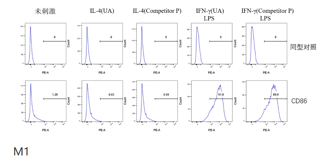
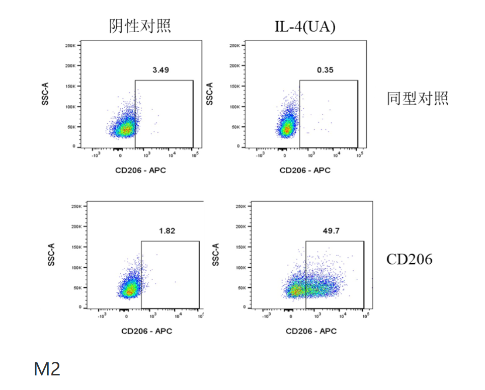
Mouse RAW264.7 Cell Polarization Induction Kit II_UA090038_优爱(UA BIOSCIENCE)官网
-
Mouse RAW264.7 Cell Polarization Induction Kit II_UA090038_ UA BIOSCIENCE Official Website
This article is reviewed and published by the technical expert team of UA
Disclaimer: This article partially utilizes artificial intelligence assistance in its creation. If any content involves copyright or intellectual property issues, please let us know and we promise to verify and remove it as soon as possible.
Purchase recombinant protein, choose Nanjing UA-Bio
UA protein focuses on providing various protein reagents, raw materials, and services required for drug research and development, cell therapy, gene therapy, and basic scientific research, including drug target proteins, immune checkpoint proteins, cytokines, tool enzymes, customized protein expression, and full-length transmembrane protein development. Youai is committed to providing customers with high-quality products and professional services, and building a High-tech Biological Enterprise with International Competitiveness.







 Business license (three certificates in one)