




In Vitro Generation of Mouse Cholangiocyte Organoids: Precise Orchestration of Core Growth Factors and Signaling Pathways


Mouse cholangiocyte organoids are miniature, self-organizing structures derived from cholangiocytes or stem cells within a three-dimensional (3D) in vitro culture system. This model effectively simulates the epithelial barrier function, secretory properties, and injury response of the bile duct. Its successful establishment and long-term maintenance are highly dependent on a precise combination of cytokines.
- Recent Advances
- Product Information
Mouse Cholangiocyte Organoids are miniature, self-organizing structures derived from cholangiocytes or stem cells in a three-dimensional in vitro culture system. This model can simulate the biliary epithelial barrier function, secretory properties, and injury response. Its successful establishment and long-term culture critically depend on a precise combination of cytokines.
Part 1: Core Functions of Key Growth Factors
The in vitro construction of cholangiocyte organoids relies on the synergistic action of several key growth factors, which provide fundamental signals for cell proliferation and morphogenesis.
-
Epidermal Growth Factor (EGF)
EGF is a potent mitogen that provides essential proliferation signals in cholangiocyte organoid culture. By binding to its receptor, EGF activates downstream signaling pathways like MAPK and PI3K-Akt, directly promoting the continuous division and expansion of cholangiocytes. This broad pro-proliferative effect supplies the necessary driving force for both initial organoid formation and subsequent passaging/expansion. -
Fibroblast Growth Factor (FGF)
Members of the Fibroblast Growth Factor family (particularly FGF10) are key factors driving the morphological patterning of cholangiocyte organoids. FGF signaling plays a central role in the embryonic development of the biliary system and, similarly in in vitro culture, guides lumen formation and budding growth in organoids. By regulating cell polarity establishment and directional migration, it promotes the generation of complex three-dimensional tubular network structures, which is crucial for building a functional bile duct model.
Part 2: Precise Regulation of Core Signaling Pathways
Beyond direct growth factors, the precise balance of several core signaling pathways is decisive for the specific differentiation and functional maintenance of cholangiocyte organoids.
-
Wnt/β-catenin Signaling Pathway
The Wnt signaling pathway is the primary switch for initiating cholangiocyte organoid formation. Moderate activation of Wnt signaling (typically mediated by the Wnt3a ligand) can induce dedifferentiation and proliferation of cholangiocytes, promoting initial organoid formation. However, the activity of this pathway must be strictly controlled at a specific level; excessive or sustained Wnt signaling activation can instead inhibit the expression of cholangiocyte-specific markers and interfere with their normal functional maturation. -
TGF-β / BMP Signaling Pathway
The Transforming Growth Factor-β and Bone Morphogenetic Protein signaling pathways form a intricate regulatory network in cholangiocyte organoids. Unlike small intestinal organoids which require strong BMP inhibition, cholangiocyte organoid culture necessitates retaining specific, moderate levels of TGF-β/BMP signaling activity. These pathways play key roles in inducing cholangiocyte-specific gene expression and establishing cell polarity. However, the balance of their concentrations is critical; overly strong signaling can induce epithelial-mesenchymal transition, leading to the disruption of organoid structure and loss of function.
Part 3: Mouse Cholangiocyte Organoid Model Construction
Preparation of Complete Medium for Mouse Cholangiocyte Organoids (Expansion): Combine basal medium, cytokines, and various additives in established proportions to prepare the complete medium.
1.Primary Culture
(1) In a biosafety cabinet, quickly dissect the mouse abdominal cavity, remove the liver tissue, and place it in pre-cooled PBS (supplemented with penicillin, streptomycin, and primary antibiotics) for washing.
(2) Collect the washed tissue fragments into a 1.5 ml EP tube. Use sterile fine-tipped ophthalmic surgical scissors to mince the tissue as much as possible (to a paste-like consistency). The degree of mincing correlates with digestion time; finer mincing shortens digestion time. Transfer the minced tissue to a 15 ml sterile centrifuge tube.
(3) Add 10 times the tissue volume of tissue digestion solution. Digest at 37°C with shaking for 10-30 minutes, monitoring progress periodically. Digestion can be terminated when a large number of cells are observed leaking out under the microscope, and most cells appear as cell clusters (Note: avoid digesting to a single-cell state).
(4) To the confirmed digested tissue suspension, add Fetal Bovine Serum (FBS) to a final concentration of 2-5%, or BSA to a final concentration of 0.1%. Mix by pipetting.
(5) Pass the tissue and cell suspension through a 70 µm cell strainer. Centrifuge the filtered cell suspension at 1000 rpm for 5 minutes. Discard the supernatant after centrifugation.
(6) If significant red pellet (red blood cells) is observed after centrifugation, add 2 ml of red blood cell lysis buffer, pipet to mix, and incubate for 3 minutes. Add 10 ml of PBS to resuspend and stop lysis. Centrifuge at 1000 rpm for 5 minutes, then discard the supernatant.
(7) Resuspend the pellet in an appropriate amount of basal medium or PBS.
(8) Mix the cell suspension with Matrigel at a suitable ratio. Using a 24-well cell culture plate as an example, plate 25-30 µl of the Matrigel-cell mixture per well (perform at 4°C).
(9) Place the plated culture plate in a 37°C incubator for 20-30 minutes to allow the Matrigel to solidify. Add an appropriate amount of pre-warmed complete medium for mouse cholangiocyte organoids (Expansion) and begin culture.
2. Organoid Passaging
(1) Aspirate the culture medium using a pipette. Add 1-2 ml of 4°C PBS per well and incubate for 2 minutes.
(2) Gently pipette to disrupt the Matrigel and collect the contents into a 15 ml centrifuge tube. Adjust the volume to 10-14 ml with PBS. Let it stand at 4°C for 20-30 minutes (pool 3-5 wells per group). Centrifuge at 1000 rpm for 5 minutes. Discard the supernatant and retain the pellet.
(3) Resuspend the organoids in an appropriate amount of fresh Matrigel. Plate 25-30 µl of the Matrigel-organoid mixture per well in a 24-well plate. Place in the incubator for 20-30 minutes for solidification. Add an appropriate amount of complete medium for mouse cholangiocyte organoids (Expansion) and continue culture.
3. Organoid Cryopreservation
(1) Aspirate the culture medium using a pipette. Add 1-2 ml of 4°C PBS per well and incubate for 2 minutes.
(2) Gently pipette to disrupt the Matrigel and collect the contents into a 15 ml centrifuge tube. Adjust the volume to 10-14 ml with PBS. Let it stand at 4°C for 20-30 minutes (pool 3-5 wells per group). Centrifuge at 1000 rpm for 5 minutes. Discard the supernatant and retain the pellet.
(3) Add an appropriate amount of organoid freezing medium. Gently pipette to resuspend. Using a 24-well plate as a reference, cryopreserve the contents of 2-3 wells per cryovial (1 ml volume per vial).
(4) Label the vials clearly. After controlled-rate freezing, transfer them to liquid nitrogen for long-term storage.
4. Organoid Thawing
(1) Pre-aliquot 10 ml of DMEM/F12 basal medium into a 15 ml centrifuge tube.
(2) Retrieve the frozen organoid vial from liquid nitrogen and quickly thaw it in a 37°C water bath.
(3) During thawing, gently agitate the cryovial to ensure complete thawing within 1-2 minutes.
(4) Quickly transfer the thawed organoid suspension to the 15 ml tube containing basal medium. Gently pipette 6-8 times. Centrifuge at 1000 rpm for 5 minutes. Discard the supernatant and collect the organoid pellet.
(5) Resuspend the pellet in Matrigel. Plate 25-30 µl of the Matrigel-organoid mixture per well in a 24-well plate. Place in the incubator for 20-30 minutes for solidification. Add an appropriate amount of complete medium for mouse cholangiocyte organoids (Expansion).
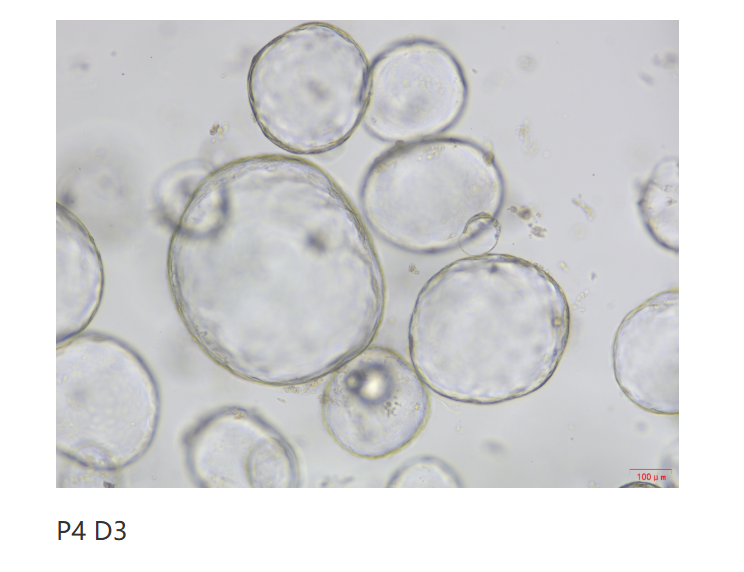
Cholangiocyte Organoid Expansion Cytokine Set, Mouse /小鼠胆管类器官(扩增)细胞因子套装_UA090044_优爱(UA BIOSCIENCE)官网
Cholangiocyte Organoid Expansion Cytokine Set, Mouse / Mouse Cholangiocyte Organoid (Expansion) Cytokine Set_UA090044_UA BIOSCIENCE Official Website
This article is reviewed and published by the technical expert team of UA
Disclaimer: This article partially utilizes artificial intelligence assistance in its creation. If any content involves copyright or intellectual property issues, please let us know and we promise to verify and remove it as soon as possible.
Purchase recombinant protein, choose Nanjing UA-Bio
UA protein focuses on providing various protein reagents, raw materials, and services required for drug research and development, cell therapy, gene therapy, and basic scientific research, including drug target proteins, immune checkpoint proteins, cytokines, tool enzymes, customized protein expression, and full-length transmembrane protein development. Youai is committed to providing customers with high-quality products and professional services, and building a High-tech Biological Enterprise with International Competitiveness.







 Business license (three certificates in one)