




A Comprehensive Guide to the Induction and Differentiation of Mouse Bone Marrow-Derived Dendritic Cells: From Cytokine Selection to Functional Assays


Dendritic cells (DCs), serving as the "sentinels" and "commanders" of the immune system, form the central bridge linking innate and adaptive immunity. Among them, bone marrow-derived dendritic cells (BMDCs) have become the gold standard model for in vitro research on DC biology, antigen presentation, T cell activation, and immune regulation, owing to their ease of large-scale acquisition and capacity for directed induction and differentiation.
- Recent Advances
- Product Information
Dendritic cells (DCs), known as the "sentinels" and "commanders" of the immune system, serve as the central bridge connecting innate and adaptive immunity. Among them, bone marrow-derived dendritic cells (BMDCs) have become the gold standard model for in vitro studies of DC biology, antigen presentation, T cell activation, and immune regulation, due to their ease of large-scale acquisition and the ability to be directionally induced to differentiate.
Faced with different research objectives—whether the need is to elicit a potent T cell response or to study immune tolerance mechanisms—selecting the correct cytokine combination is key to success. This guide will simplify the complex, providing an in-depth analysis of how to use cytokine kits to efficiently induce functionally specific mouse BMDCs in vitro.
Part 1: Theoretical Foundation – Understanding the Two Major Differentiation Pathways of BMDCs
Mouse bone marrow precursor cells can primarily be induced to differentiate into two major classes of DCs with distinct functions in vitro:
-
Conventional DCs (cDCs): Driven to differentiate by GM-CSF. These cells highly express MHC class II molecules and co-stimulatory molecules (CD80, CD86), possess strong antigen-presenting capacity, and can effectively activate naïve T cells. They are the most commonly used model for studying inflammation, infection, and anti-cancer immunity.
-
Plasmacytoid DCs (pDCs): Driven to differentiate by Flt3 Ligand. These cells are characterized by massive production of Type I interferons and play a central role in antiviral immunity and certain autoimmune diseases.
Part 2: Core Arsenal – Core Signaling Pathways Driving BMDC Differentiation
Cytokines themselves are the "messengers," while signaling pathways are the "command execution systems" within the cell. The following are the most critical pathways in inducing BMDCs:
-
JAK-STAT Signaling Pathway
This is the core pathway through which most cytokines function.-
Mode of Action: Cytokines (such as GM-CSF, IL-4, Flt3-L) bind to receptors on the cell surface, causing conformational changes in the receptor and activating the associated JAK kinases. The activated JAKs phosphorylate the receptor, creating docking sites for STAT proteins. The phosphorylated STAT proteins form dimers, translocate into the nucleus, and act as transcription factors to directly regulate the expression of specific genes.
-
Example: GM-CSF primarily drives myeloid precursor cells toward cDC differentiation via the JAK2/STAT5 pathway.
-
-
NF-κB Signaling Pathway
This is the "master switch" for DC maturation.-
Mode of Action: Maturation stimuli (such as LPS via TLR4, TNF-α via its receptor) activate the IKK complex, which subsequently phosphorylates and degrades IκB proteins, allowing NF-κB dimers (e.g., p65/p50) to be released and enter the nucleus. This initiates the transcription of a large number of pro-inflammatory cytokines (TNF-α, IL-6, IL-12), chemokines, and co-stimulatory molecules (CD80, CD86).
-
-
MAPK Signaling Pathway
Cooperatively regulates cell proliferation, survival, and inflammatory responses.-
Mode of Action: Also activated by various stimuli (including growth factors, stress, LPS), it includes branches such as ERK, JNK, and p38. In DCs, the p38 MAPK pathway is crucial for IL-12 production.
-
Part 3: Practical Protocol – Mouse BMDC Induction and Differentiation Protocol
1. Isolation of Mouse Bone Marrow Cells
1.1 Euthanize 6-8 week-old mice by cervical dislocation. Harvest femurs and remove surrounding muscle tissue.
1.2 Disinfect femurs by immersing in 75% ethanol for 2 minutes, then wash twice with PBS.
1.3 Use scissors to cut off both ends of the femurs. Flush the bone marrow cavity repeatedly with PBS using a syringe and needle inserted into the ends until the bone appears white.
1.4 Collect the bone marrow suspension and filter it through a 200-mesh nylon filter.
1.5 Centrifuge the filtrate at 1200 rpm for 5 minutes. Discard the supernatant.
1.6 Add 2 ml of red blood cell lysis buffer and incubate at room temperature for 3-5 minutes.
1.7 Add 10 ml of PBS and centrifuge at 1200 rpm for 5 minutes.
1.8 Discard the supernatant, wash the cell pellet once with PBS, and resuspend the cells in 1640 medium supplemented with 10% FBS.
2. BMDC Induction
2.1 Adjust the cell density to 1x10^6/mL using the medium. Add GM-CSF (UA040056) (20 ng/mL) and IL-4 (UA040192) (20 ng/mL). This is designated Day 0 (D0).
2.2 Perform a semi-medium change every 2 days: Remove half of the medium, centrifuge to collect the suspended cells, resuspend them in an equal volume of fresh medium supplemented with the corresponding cytokines, and add back to the dish.
2.3 On D6, collect both suspended and loosely adherent cells. Centrifuge at 1200 rpm for 5 minutes, discard the supernatant. Adjust the cell density to 1x10^6/mL and add GM-CSF (20 ng/mL) and IL-4 (20 ng/mL). These are incompletely mature BMDCs.
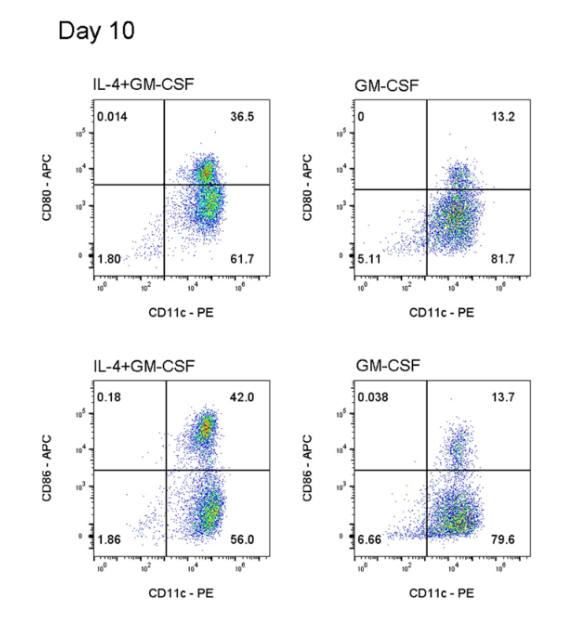
2.4 Full Maturation of BMDCs: On D8, add TNF-α (20 ng/mL), GM-CSF (20 ng/mL), and IL-4 (20 ng/mL). On D10, collect both suspended and adherent cells.
3. Detection of Incompletely Mature BMDCs
3.1 Cell Collection: After cytokine treatment, discard the old medium, wash the cells once with PBS, discard the supernatant, and then gently detach adherent cells using PBS and resuspend.
3.2 Counting: Count the cells using a cell counter, calculate the total cell number, and adjust the concentration to 1x10^7/mL.
3.3 Cell Blocking: Add 100 µl/well to a 96-well plate or flow cytometry tube. Add Mouse IgG (Mouse FcR Blocking Reagent). Incubate at 4°C for 30 min. Centrifuge at 300 x g for 5 min. Discard supernatant.
3.4 Antibody Staining: Add the following antibodies to respective tubes/groups. Incubate at 4°C for 30 min, protected from light. Centrifuge at 300 x g for 5 min. Discard supernatant.
* Group 1: PE anti-mouse CD11c Antibody (117307) (Refer to datasheet for amount)
APC anti-mouse CD80 Antibody (104713) (Refer to datasheet for amount)
* Group 2: PE anti-mouse CD11c Antibody (117307) (Refer to datasheet for amount)
APC anti-mouse CD86 Antibody (105011) (Refer to datasheet for amount)
3.5 Cell Washing: Wash cells with PBS to remove residual antibody, and resuspend in PBS.
3.6 Viability Staining: Add 7-AAD (559925) to each tube/well. Incubate at room temperature for 5 min, protected from light.
3.7 Flow Cytometry Analysis.
4. Detection of Fully Mature BMDCs
4.1 Cell Collection: After cytokine treatment, discard the old medium, wash the cells once with PBS, discard the supernatant, and then gently detach adherent cells using PBS and resuspend.
4.2 Counting: Count the cells using a cell counter, calculate the total cell number, and adjust the concentration to 1x10^7/mL.
4.3 Cell Blocking: Add 100 µl/well to a 96-well plate or flow cytometry tube. Add Mouse IgG (Mouse FcR Blocking Reagent). Incubate at 4°C for 30 min. Centrifuge at 300 x g for 5 min. Discard supernatant.
4.4 Antibody Staining: Add the following antibodies to respective tubes/groups. Incubate at 4°C for 30 min, protected from light. Centrifuge at 300 x g for 5 min. Discard supernatant.
* Group 1: PE anti-mouse CD11c Antibody (117307) (Refer to datasheet for amount)
APC anti-mouse CD80 Antibody (104713) (Refer to datasheet for amount)
* Group 2: PE anti-mouse CD11c Antibody (117307) (Refer to datasheet for amount)
APC anti-mouse CD86 Antibody (105011) (Refer to datasheet for amount)
4.5 Cell Washing: Wash cells with PBS to remove residual antibody, and resuspend in PBS.
4.6 Viability Staining: Add 7-AAD (559925) to each tube/well. Incubate at room temperature for 5 min, protected from light.
4.7 Flow Cytometry Analysis.

This article is reviewed and published by the technical expert team of UA
Disclaimer: This article partially utilizes artificial intelligence assistance in its creation. If any content involves copyright or intellectual property issues, please let us know and we promise to verify and remove it as soon as possible.
Purchase recombinant protein, choose Nanjing UA-Bio
UA protein focuses on providing various protein reagents, raw materials, and services required for drug research and development, cell therapy, gene therapy, and basic scientific research, including drug target proteins, immune checkpoint proteins, cytokines, tool enzymes, customized protein expression, and full-length transmembrane protein development. Youai is committed to providing customers with high-quality products and professional services, and building a High-tech Biological Enterprise with International Competitiveness.







 Business license (three certificates in one)