




Unveiling the Intelligent Probe for Intracellular Dynamics: pH-sensitive IgG Labeling Reagents Max(Green)


pH-sensitive IgG labeling reagents Max(Green) are specially designed chemical reagents for covalently labeling antibodies (IgG).
- Recent Advances
- Product Information
At the forefront of life science research, scientists are no longer satisfied with merely knowing "where" a protein is; they are eager to understand "what it is experiencing" inside the cell. This is especially true for large molecules like antibodies, which, after being engulfed by cells, undergo complex transport and fate decisions within various vesicles. During this process, changes in environmental acidity/alkalinity (pH) are key signals. pH-sensitive IgG Labeling Reagents Max(Green) is a powerful tool developed in response to this need. It acts like an "intelligent tracker," capable of precisely labeling antibodies while simultaneously reporting the pH changes at their location in real-time, thereby revealing the dynamic life activities within cells.
I. pH-sensitive IgG Labeling Reagents Max(Green): A Dual-Purpose Tool for Precise Localization and Environmental Sensing
pH-sensitive IgG Labeling Reagents Max(Green) are specially designed chemical reagents used for covalently labeling antibodies (IgG). Its name contains three key pieces of information:
pH-sensitive: This is its core characteristic, meaning its optical properties (such as fluorescence intensity) change with the pH of the environment.
IgG Labeling: It contains chemical groups that can efficiently and stably bind to specific amino acids (e.g., lysine) on antibody molecules (Immunoglobulin G).
Max(Green):
Max: Represents maximized labeling efficiency. Through optimized reaction conditions, it can attach as many fluorescent dye molecules as possible to each antibody molecule, resulting in a very strong fluorescent signal and greatly enhancing detection sensitivity.
Green: Indicates it emits green fluorescence, making it suitable for conventional fluorescence microscopes and flow cytometers equipped with FITC or GFP filter sets.
In short, it is a reagent that can turn an antibody into an intelligent pH sensor.
II. Mechanism of Action -- Fluorescence Intensity "Dances" with pH
Its core working principle is based on the pH-dependent fluorescence quenching effect of the dye.
In Neutral/Alkaline Environments: When the labeled antibody is in the extracellular fluid (pH ~7.4) or in compartments with higher pH like early endosomes, the dye molecule is in a deprotonated state. This state allows the dye to efficiently emit bright green fluorescence upon laser excitation.
In Acidic Environments: When the antibody is endocytosed and enters progressively acidifying endosomes and lysosomes (pH dropping from 6.5 to 4.5), the dye molecule binds protons. This protonation causes a change in the electronic structure of the dye, greatly inhibiting fluorescence emission, leading to a significant weakening or even complete quenching of the fluorescent signal.
This process is usually reversible. If the vesicle's pH increases again, the fluorescence intensity can recover. This property allows researchers to dynamically observe cyclical pH changes.
III. Main Applications -- Visualizing Cellular "Logistics" and "Digestion" in Real-Time
Leveraging its unique mechanism, Max(Green) plays an irreplaceable role in the following research areas:
1. Study of Antibody Drug Endocytosis and Intracellular Trafficking Pathways
This is its most classic and important application. In antibody drug development, understanding how a drug enters the cell, where it goes, and when it is degraded is crucial.
Experimental Workflow: Label the candidate therapeutic antibody with Max(Green).
Observation Process:
Binding Stage: The antibody binds to the cell surface, emitting strong green fluorescence.
Endocytosis & Transport Stage: The antibody enters the cell, moving from early endosomes (slightly lower pH, fluorescence begins to weaken) to late endosomes (even lower pH, fluorescence weaker).
Degradation Stage: The antibody finally arrives in lysosomes (pH ~4.5, highly acidic), where fluorescence is almost completely quenched.
Research Value: By tracking fluorescence intensity changes in real-time, one can precisely quantify the endocytosis rate, transport kinetics, and the time taken to reach lysosomes, providing critical data for optimizing antibody drug delivery and efficacy.
2. Receptor-Mediated Endocytosis and Signal Transduction
Many receptors on the cell membrane (e.g., growth factor receptors, G protein-coupled receptors) initiate endocytosis after binding their ligands.
Research Method: Use Max(Green) to label specific antibodies against the receptor to track the internalization and recycling process of the receptor itself.
Research Value: Reveals how receptors are regulated (e.g., whether they enter the degradation pathway to terminate signaling or recycle back to the membrane for reuse). This is crucial for understanding aberrant signaling pathways in diseases like cancer.
3. Diagnosis of Lysosomal Function and Related Diseases
Lysosomes are the "digestive organs" of the cell, and their acidic environment is key to their normal function.
Research Method: Using probes labeled with Max(Green) (e.g., antibodies targeting lysosomal membrane proteins) can indirectly assess lysosomal pH homeostasis.
Research Value: If fluorescence persists within lysosomes, it suggests insufficient lysosomal acidification and functional impairment. This is closely associated with various neurodegenerative diseases (e.g., Alzheimer's), metabolic diseases, and lysosomal storage disorders.
4. Research on Viral and Pathogen Invasion Mechanisms
Many viruses and bacterial toxins utilize the endocytic pathway for cell entry, triggering the fusion of their envelope with the host cell membrane in the acidic environment.
Research Method: Labeling antibodies targeting viral or bacterial surface proteins with Max(Green) allows simulation and visualization of the entire entry path.
Research Value: Helps elucidate the invasion mechanisms of pathogens, providing new targets for developing drugs that block infection.
IV. How to Use pH-sensitive IgG Labeling Reagents Max(Green)
Using UA BIOSCIENCE's pH-sensitive IgG Labeling Reagents Max(Green) UA070122 as an example:
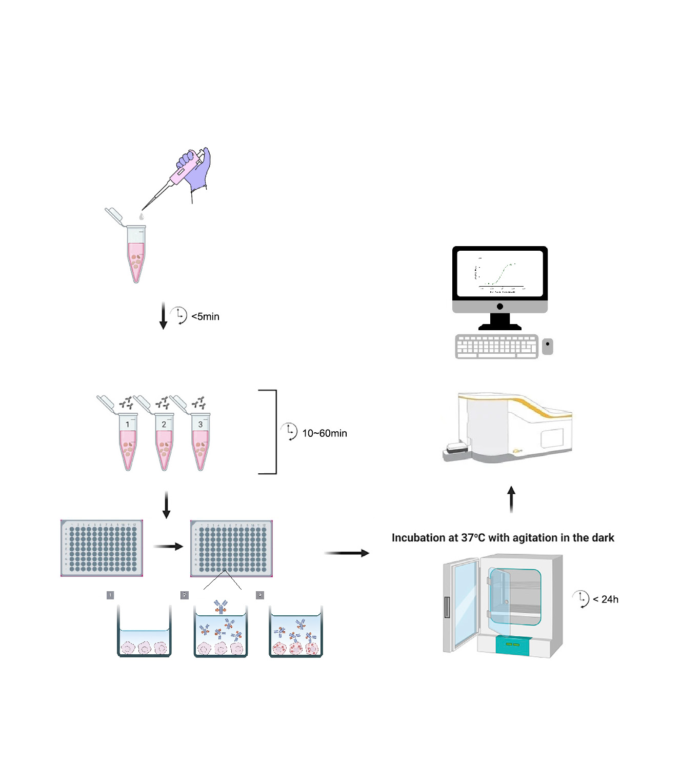
1. Incubation of UA070122 with Antibody
1.1. Reconstitute the lyophilized UA070122 powder using deionized water according to the dissolution instructions.
1.2. Prepare a sufficient volume of a 4X working solution of the test antibody in cell culture medium (four times the final test concentration, determined based on prior flow cytometry optimization). For example, 2 µg/ml might be a good starting test concentration for the antibody, so the 4X working solution would be 8 µg/ml.
1.3. Prepare a sufficient volume of a 4X working solution of UA070122 in cell culture medium. Use a molar ratio of test antibody to UA070122 of 1:2.
1.4. Mix the 4X test antibody working solution and the 4X UA070122 working solution in a 1:1 volume ratio. Incubate at room temperature, protected from light, for 15 minutes to 1 hour, to obtain the Ab-UA070122 complex 2X working solution.
2. Incubation of Ab-UA070122 Complex with Cells
2.1. Cell Preparation: Harvest and wash cells. Adjust cell concentration using complete medium. Suspend cells at 1-2×10^5 cells/mL for suspension cells, or seed adherent cells at 0.5-1×10^5 cells/mL, adding 100 µL of cell suspension per well of a 96-well plate.
2.2. Incubation: Add 100 µL of the Ab-UA070122 complex 2X working solution to each well. Culture in a 37°C, 5% CO2 incubator.
3. Flow Cytometry Detection
After culturing for 18-24 hours (adjustable based on the actual endocytosis kinetics of the antibody), harvest the cells and analyze the endocytosis effect of the antibody by flow cytometry. Use the FITC or AF488 channel for detection.
pH-sensitive IgG Labeling Reagents Max(Green)_UA070122_UA BIOSCIENCE Official Website
This article is reviewed and published by the technical expert team of UA
Disclaimer: This article partially utilizes artificial intelligence assistance in its creation. If any content involves copyright or intellectual property issues, please let us know and we promise to verify and remove it as soon as possible.
Purchase recombinant protein, choose Nanjing UA-Bio
UA protein focuses on providing various protein reagents, raw materials, and services required for drug research and development, cell therapy, gene therapy, and basic scientific research, including drug target proteins, immune checkpoint proteins, cytokines, tool enzymes, customized protein expression, and full-length transmembrane protein development. Youai is committed to providing customers with high-quality products and professional services, and building a High-tech Biological Enterprise with International Competitiveness.







 Business license (three certificates in one)