




Annexin V-FITC/7-AAD Apoptosis Detection Kit Principle and Applications


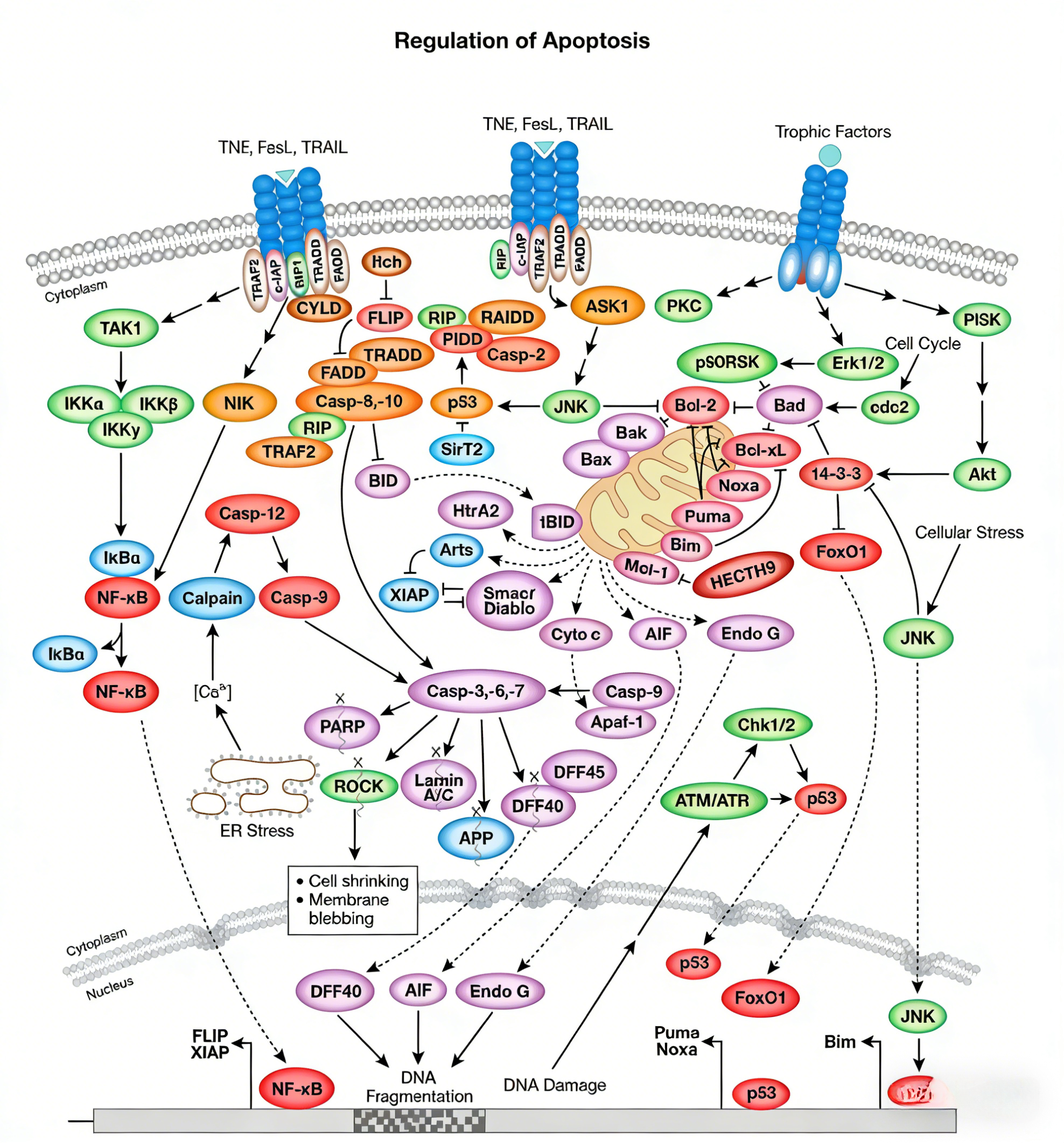
Apoptosis is a highly conserved form of programmed cell death in organisms, playing a crucial role in various physiological and pathological processes such as embryonic development, tissue homeostasis maintenance, immune system regulation, and disease progression.
- Recent Advances
I. Biological Significance of Apoptosis Detection
Apoptosis is a highly conserved programmed cell death mechanism in organisms, playing a crucial role in various physiological and pathological processes such as embryonic development, tissue homeostasis maintenance, immune system regulation, and disease progression. Dysregulated apoptosis is closely associated with numerous diseases, including cancer, autoimmune disorders, neurodegenerative diseases, and infectious diseases. Therefore, accurate and sensitive detection and quantification of apoptosis are indispensable for fundamental life science research, drug screening, and toxicological evaluation.
II. Core Principle of Annexin V-FITC/7-AAD Detection Method
This detection method is based on two early key events during apoptosis—phosphatidylserine externalization and loss of membrane integrity—enabling precise discrimination among viable cells, early apoptotic cells, and late apoptotic/necrotic cells.
1. Annexin V-FITC Identifies Early Apoptosis:
In normal viable cells, phosphatidylserine (PS) is predominantly located on the inner leaflet of the plasma membrane. During early apoptosis, PS rapidly and specifically translocates to the outer membrane, serving as an "eat me" signal for phagocytes to recognize and clear apoptotic cells. Annexin V is a calcium-dependent phospholipid-binding protein with high affinity and specificity for PS. The Annexin V conjugated to the green fluorescent dye FITC in the kit binds to PS on the outer membrane of apoptotic cells, allowing sensitive detection of early apoptotic cells via flow cytometry or fluorescence microscopy.
2. 7-AAD Distinguishes Membrane Integrity:
7-Aminoactinomycin D (7-AAD) is a nucleic acid dye that cannot penetrate intact cell membranes, thus failing to stain viable or early apoptotic cells. When cells enter late apoptosis or necrosis, membrane integrity is compromised, allowing 7-AAD to enter cells and bind DNA, emitting intense red fluorescence. Therefore, 7-AAD fluorescence detection effectively distinguishes cells with intact membranes (Annexin V⁺/7-AAD⁻ for early apoptosis) from those with compromised membranes (Annexin V⁺/7-AAD⁺ for late apoptosis/necrosis; Annexin V⁻/7-AAD⁺ typically indicates necrotic cells).
III. Core Advantages and Technical Features of the Kit
1. High Sensitivity and Specificity: The high specificity of Annexin V for PS ensures accurate detection of early apoptosis, avoiding nonspecific signal interference.
2. Clear Subpopulation Discrimination: Dual-parameter analysis of FITC and 7-AAD clearly divides cell populations into four quadrants: viable cells (double-negative), early apoptotic cells (FITC⁺/7-AAD⁻), late apoptotic/necrotic cells (FITC⁺/7-AAD⁺), and necrotic cells (FITC⁻/7-AAD⁺), enabling precise quantification.
3. Simple and Rapid Operation: The kit typically provides ready-to-use binding buffer and optimized working solution formulations, featuring simple staining steps and short incubation times (usually 15–20 minutes). It is compatible with flow cytometry and fluorescence microscopy platforms for rapid results.
4. Applicability to Diverse Sample Types: Widely suitable for apoptosis detection in suspension cells (e.g., lymphocytes, tumor cell lines) and adherent cells (after gentle digestion), as well as certain primary cells.
5. Live-Cell Analysis Capability: Compared to traditional dyes like propidium iodide, 7-AAD exhibits lower toxicity toward early apoptotic cells, facilitating live-cell analysis.
IV. Major Application Areas
1. Fundamental Cell Biology Research: Investigating apoptosis mechanisms and signaling pathways induced by various stimuli, such as growth factor deprivation, drug treatment, radiation, and hypoxia.
2. Anticancer Drug Screening and Efficacy Evaluation: High-throughput screening of candidate compounds with apoptosis-inducing activity and assessing their efficacy in vitro and in preclinical models.
3. Immunology Research: Detecting activation-induced apoptosis in immune cells (e.g., T cells, B cells) and studying mechanisms of immune tolerance and autoimmune diseases.
4. Toxicology and Safety Assessment: Evaluating potential toxicity of environmental toxins, chemical drugs, or nanomaterials and their apoptosis-inducing capacity.
5. Stem Cell and Developmental Biology: Studying apoptotic events during stem cell differentiation, tissue development, or degeneration.
V. Standard Experimental Protocol and Key Considerations
1. Sample Preparation: Harvest cells, wash with pre-cooled PBS, and resuspend in 1X binding buffer at an appropriate cell density (typically 10⁵–10⁶ cells/mL).
2. Staining Incubation: Add Annexin V-FITC and 7-AAD working solutions to the cell suspension, mix gently, and incubate at room temperature (20–25°C) in the dark for 15–20 minutes.
3. Instrument Detection: After incubation, analyze immediately (preferably within 1 hour) using a flow cytometer. Set appropriate fluorescence channels (FITC: FL1; 7-AAD: FL3 or equivalent) and adjust fluorescence compensation using unstained and single-stained controls.
4. Key Considerations:
- Avoid buffers containing EDTA or digestive enzymes (e.g., trypsin), as they may interfere with Annexin V-calcium binding or damage cell membranes.
- Handle cells gently to prevent mechanical damage and false positives.
- Perform detection promptly after incubation to avoid changes in cell status over time.
- For adherent cells, use gentle digestion solutions without EDTA.
- Include appropriate positive and negative controls (e.g., apoptosis-inducer-treated cells) for accurate data interpretation.
VI. Summary and Outlook
The Annexin V-FITC/7-AAD apoptosis detection kit, with its detection principle based on early apoptotic events, clear cell population discrimination, and operational convenience, has become one of the most widely used and reliable gold-standard methods in apoptosis research. It provides a powerful quantitative tool for life science research and drug development. In the future, with advancements in multicolor flow cytometry and imaging technologies, this kit can be combined with additional cellular markers (e.g., cell cycle, reactive oxygen species, mitochondrial membrane potential) for multiparameter analysis, enabling comprehensive exploration of cell fate determination networks at the single-cell level and advancing precision medicine and translational research.
Disclaimer: This article partially utilizes artificial intelligence assistance in its creation. If any content involves copyright or intellectual property issues, please let us know and we promise to verify and remove it as soon as possible.
Purchase recombinant protein, choose Nanjing UA-Bio
UA protein focuses on providing various protein reagents, raw materials, and services required for drug research and development, cell therapy, gene therapy, and basic scientific research, including drug target proteins, immune checkpoint proteins, cytokines, tool enzymes, customized protein expression, and full-length transmembrane protein development. Youai is committed to providing customers with high-quality products and professional services, and building a High-tech Biological Enterprise with International Competitiveness.







 Business license (three certificates in one)