




Study on TNF-α signaling pathway and the therapeutic mechanisms of its inhibitors


Tumor necrosis factor α (TNF-α) is a pleiotropic cytokine composed of 157 amino acids that functions as a homotrimer.
- Recent Advances
- Product Information
I. Molecular Structure and Biological Characteristics of TNF-α
Tumor necrosis factor alpha (TNF-α) is a cytokine with pleiotropic biological effects, composed of 157 amino acids that functions as a homotrimer. It is primarily produced by activated macrophages, T lymphocytes, and natural killer cells, and can induce the expression of various inflammatory molecules (including cytokines and chemokines), occupying a central position in the regulation of inflammatory responses.
TNF-α exists in two forms: transmembrane TNF-α (tmTNF-α) and soluble TNF-α (sTNF-α). tmTNF-α is the initially synthesized precursor form, which requires processing by TNF-α converting enzyme (TACE) to release sTNF-α. The two forms mediate biological effects by binding to different receptors: sTNF-α primarily acts on TNFR1, while tmTNF-α can bind to both receptors but its biological activity is mainly mediated through TNFR2. In related mechanism studies, the Mouse TNF-α Kit (HICA) is widely used for quantitative detection of TNF-α levels in animal model samples, providing an important tool for analyzing its in vivo functions.
II. TNF-α Receptor System and Signal Transduction Mechanisms
(1) Tissue Distribution and Structural Features of Receptors
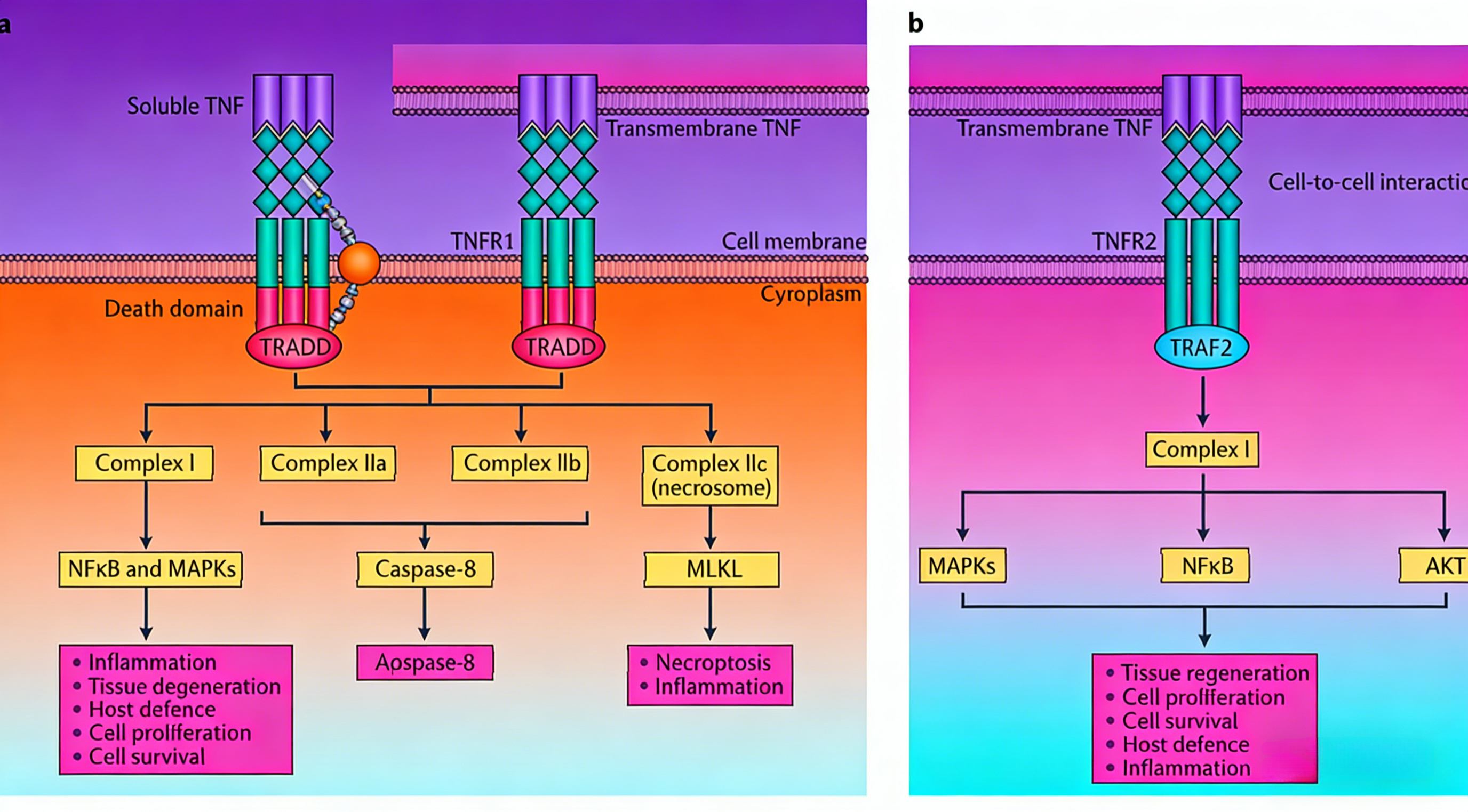
TNF-α exerts diverse biological functions by binding to two different receptors. TNFR1 is widely expressed in all human tissues, and its intracellular domain contains a conserved death domain, making it a key signaling receptor for TNF-α-mediated inflammation and cell death. TNFR2 expression is limited to specific cell types, including neurons, immune cells, and endothelial cells. This receptor lacks a death domain and cannot directly induce apoptosis; it is primarily activated by tmTNF-α.
(2) Assembly of Signaling Complexes After Receptor Activation
Signal transduction is initiated when the TNF homotrimer binds to the TNFR homotrimer. After TNFR1 activation, it recruits TNFR1-associated death domain protein (TRADD) to assemble different signaling complexes:
Complex I assembles at the plasma membrane and includes components such as TRADD, RIPK1, TRAF2/5, cIAP1/2, and LUBAC. It activates the NF-κB and MAPK signaling pathways, inducing the expression of inflammation-related genes and playing a crucial role in host defense, cell proliferation, and survival.
Complexes IIa, IIb, and IIc assemble in the cytoplasm. Complex IIa consists of TRADD, RIPK1, TRAF2, cIAP1/2, pro-Caspase-8, and FADD; Complex IIb adds RIPK3 to this composition. Together, they are referred to as the apoptosome and induce apoptosis through caspase-8 activation. Complex IIc (the necrosome) is formed by the binding of RIPK1 and RIPK3, which activate mixed lineage kinase domain-like protein (MLKL) through RIPK3-mediated phosphorylation, inducing necroptosis and inflammatory responses.
TNFR1 primarily promotes inflammation and tissue degeneration, while TNFR2 mediates local homeostatic effects, including cell survival and tissue regeneration.
III. Physiological Functions and Pathological Significance of TNF-α
Under physiological conditions, TNF-α is a key component of normal immune responses, participating in host defense by activating the immune system. However, inappropriate or excessive production of TNF-α can lead to pathological damage and is closely associated with the pathogenesis of various autoimmune diseases.
Clinical studies have confirmed that abnormal TNF-α secretion is involved in the development of the following diseases: rheumatoid arthritis (RA), inflammatory bowel disease (IBD), psoriatic arthritis (PsA), ankylosing spondylitis (AS), psoriasis (PS), and non-infectious uveitis (NIU). In these disease states, persistent overproduction of TNF-α drives chronic inflammatory responses and tissue damage. In animal model studies, the Mouse TNF-α Kit (HICA) is used to detect changes in cytokine levels in disease models, providing data support to elucidate the role of TNF-α in pathogenesis.
IV. Mechanism of Action and Therapeutic Value of TNF-α Inhibitors
Given the central pathogenic role of TNF-α in autoimmune diseases, targeting TNF-α has become an important therapeutic strategy. TNF-α inhibitors specifically bind to TNF-α, blocking its interaction with cell surface TNFR1 and TNFR2, thereby inhibiting the activation of downstream inflammatory signaling pathways.
The therapeutic value of these drugs has been validated in various autoimmune diseases. By neutralizing excess TNF-α, they effectively reduce inflammatory responses, delay tissue damage, and improve patients' clinical symptoms and quality of life. In preclinical studies, the Mouse TNF-α Kit (HICA) is used to evaluate the inhibitory effects of candidate drugs on TNF-α levels in animal models, providing critical data for drug screening and dose optimization.
V. Summary and Future Perspectives
As a core regulator of inflammatory responses, the molecular structure, receptor system, and signal transduction mechanisms of TNF-α have been extensively studied. The fine-tuned regulation of two receptors and multiple signaling complexes determines the diversity of TNF-α functions—ranging from physiological immune defense to pathological inflammatory damage. TNF-α inhibitors, by blocking key nodes in this pathway, provide effective treatment for autoimmune diseases.
Future research could further explore the spatiotemporal specificity of TNF-α signaling networks and develop more selective intervention strategies. Meanwhile, detection tools like the Mouse TNF-α Kit (HICA) will continue to play a vital role in basic research, animal model validation, and preclinical efficacy evaluation, providing technical support for a deeper understanding of TNF-α's biological functions and the optimization of therapeutic strategies.
VI. Which Manufacturers Provide the Mouse TNF-α Kit (HICA)?
Nanjing UA-Bio Technology Co., Ltd. (UA-Bio) has independently developed the "Mouse TNF-α Kit (HICA)", a high-performance in vitro detection platform specifically designed for studying key pathways in murine inflammatory responses and apoptosis. This kit is engineered to accurately and efficiently quantify the immunobinding activity of mouse tumor necrosis factor-alpha (TNF-α) protein, offering a stable and reliable standardized solution for mechanistic research and preclinical drug efficacy analysis in fields such as autoimmune diseases, inflammatory disorders, tumor immunology, and more.
| Core Advantages of the Product |
|---|
| High Purity and Complete Biological Activity: The core components of the kit utilize high-purity, high-biological-activity mouse-derived TNF-α protein validated through multi-dimensional quality control. This protein maintains the correct native homotrimeric conformation and full receptor-binding capacity, faithfully simulating the inflammatory and apoptotic signals mediated by mouse TNF-α under physiological conditions, ensuring the accuracy, reproducibility, and functional relevance of binding assay data. |
| Exceptional Batch-to-Batch Consistency and Stability: Leveraging an internationally leading recombinant protein expression platform and highly standardized purification processes, combined with a rigorous release quality control system, the product exhibits outstanding long-term stability and excellent batch-to-batch consistency. This provides solid and reliable quality assurance for long-term, continuous preclinical research and high-throughput screening. |
| Ready-to-Use Flexible Detection Platform: Based on an optimized enzyme-linked immunosorbent assay (ELISA) principle, the kit provides pre-coated strips, highly specific detection antibodies, standards, and a complete set of optimized buffer systems. It features simple and rapid operation, high sensitivity, and strong specificity, making it widely applicable for various research needs, including anti-mouse TNF-α antibody/receptor antagonist screening, neutralizing activity determination, competitive binding assays, affinity analysis, and immunogenicity evaluation. |
| Comprehensive Solutions and Professional Support: We provide thoroughly validated standard protocols, typical dose-response curves, and detailed result interpretation guides to help you quickly establish stable and reproducible detection workflows. Nanjing UA-Bio's professional technical team offers full-process, expert technical consultation and support for your research design, experimental optimization, and data analysis. |
Nanjing UA-Bio Technology Co., Ltd. is committed to providing cutting-edge, high-quality core reagents and tools for immunology, cell therapy, and innovative drug development. For detailed technical specifications, validation data, or specific application inquiries regarding the "Mouse TNF-α Kit (HICA)" (Catalog No.: UA086048), please feel free to contact us.
This article is reviewed and published by the technical expert team of UA
Disclaimer: This article partially utilizes artificial intelligence assistance in its creation. If any content involves copyright or intellectual property issues, please let us know and we promise to verify and remove it as soon as possible.
Purchase recombinant protein, choose Nanjing UA-Bio
UA protein focuses on providing various protein reagents, raw materials, and services required for drug research and development, cell therapy, gene therapy, and basic scientific research, including drug target proteins, immune checkpoint proteins, cytokines, tool enzymes, customized protein expression, and full-length transmembrane protein development. Youai is committed to providing customers with high-quality products and professional services, and building a High-tech Biological Enterprise with International Competitiveness.







 Business license (three certificates in one)