




Development and Application Research of pH-Sensitive IgG Labeling Reagents


Immunofluorescence labeling technology is an indispensable tool in modern biomedical research, where immunoglobulin G (IgG), as the most commonly used secondary or primary antibody carrier, directly influences the sensitivity and specificity of detection.
- Recent Advances
- Product Information
I. Introduction
Immunofluorescence labeling technology is an indispensable tool in modern biomedical research. Immunoglobulin G (IgG), as the most commonly used secondary or primary antibody carrier, directly influences the sensitivity and specificity of detection through its labeling strategy. Traditional labeling reagents lack responsiveness to environmental changes in complex physiological environments, particularly in dynamic processes involving pH changes such as endocytosis and lysosomal degradation, making it difficult to distinguish between antibody binding on the cell surface and acidification accumulation within endosomes.
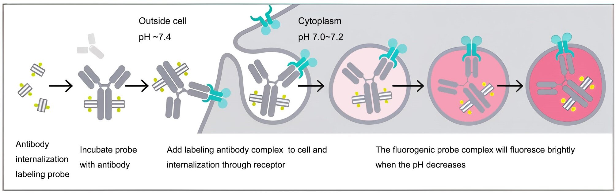
The emergence of pH-sensitive fluorescent dyes provides a novel solution to these challenges. These dyes exhibit weak fluorescence signals under physiological conditions (pH 7.2-7.4) but show significantly enhanced fluorescence intensity in acidic environments (pH 4.5-6.0). By conjugating such dyes with IgG, an "intelligent" probe can be constructed to monitor the transport pathway and acidification process of antibodies after entering cells in real-time and dynamically. Among these, dyes with emission wavelengths in the red light region are preferred for live imaging and tissue section analysis due to their strong tissue penetration and low autofluorescence interference.
II. Reagent Design and Mechanism of Action
2.1 Molecular Structural Features
pH-sensitive red labeling reagents are typically designed based on the intramolecular spirocyclic structure of rhodamine or its derivatives. In molecular design, the introduction of protonatable nitrogen atoms or specific spirocyclic switches on the fluorescent core enables responsiveness to hydrogen ion concentration. In neutral environments, the molecule remains in a spiro-closed state with an interrupted conjugated system, resulting in fluorescence quenching. In acidic environments, the spiro ring opens, forming an oxazine structure with a large conjugated plane, thereby releasing intense red fluorescence.
2.2 Spectral Characteristics of pH Response
The maximum excitation wavelength of this reagent typically ranges between 560-580 nm, and the maximum emission wavelength is between 590-610 nm, falling within the standard "red" spectral window. Its key pH response range (pKa value) is precisely regulated, mainly between 5.0 and 6.5, which highly matches the acidification gradient of intracellular endosomes (pH 6.0-6.5) and lysosomes (pH 4.5-5.5). At pH 7.4, the fluorescence intensity is only one-tenth to one-twentieth of that at pH 5.0, demonstrating an extremely high signal-to-noise ratio.
III. IgG Labeling Methods and Process Optimization
3.1 Conjugation Chemistry Principles
Efficient labeling requires the use of mild and stable bioconjugation techniques. Typically, pH-sensitive dyes containing N-hydroxysuccinimide (NHS) ester-activated groups are employed. NHS esters can specifically react with primary amines on lysine residues of IgG molecules to form stable amide bonds. This reaction is gentle and can be completed within a few hours of incubation at room temperature or 4°C in a sodium bicarbonate buffer (pH 8.3-8.5).
3.2 Labeling Efficiency and Quality Control
The molar ratio of dye to antibody (F/P ratio) must be strictly controlled during labeling. An excessively high F/P ratio may alter antibody conformation or increase steric hindrance at antigen-binding sites, while an excessively low F/P ratio may reduce imaging sensitivity. Experimental optimization suggests an F/P ratio between 2 and 4. Post-labeling, the product should be purified via gel filtration chromatography to remove free dye, and protein concentration and dye binding rate should be determined using UV-visible spectrophotometry to ensure batch-to-batch consistency.
IV. Experimental Characterization and Performance Validation
4.1 In Vitro pH Response Curve Determination
The labeled IgG is diluted in phosphate buffers of varying pH (4.0 to 8.0), and fluorescence intensity changes are measured using a fluorescence microplate reader. Results show that fluorescence intensity increases in a typical "S"-shaped curve as pH decreases. At pH 6.0, fluorescence intensity is approximately 15 times higher than at pH 7.4, confirming the reagent's excellent "light-up" characteristics in simulated endosomal environments.
4.2 Targeted Binding and Endocytosis Imaging
Immunofluorescence staining is performed using cell lines expressing specific membrane antigens. At 4°C staining conditions, only weak fluorescence signals are observed on the cell membrane. When the temperature is raised to 37°C to induce endocytosis, punctate bright red fluorescence appears intracellularly over time (15 minutes to 2 hours) and gradually aggregates around the nuclear region. This result visually demonstrates the complete dynamic process of antibody binding, internalization, and eventual entry into acidic organelles—a distinction unattainable with conventional labeling dyes.
V. Application Scenarios Analysis
5.1 Receptor-Mediated Endocytosis Mechanism Research
In neuroscience, using this labeling reagent to track the transport trajectory of neurotrophic factor receptors can distinguish between receptors in the signaling-active phase (membrane-bound) and the degradation phase (within lysosomes), providing direct evidence for deciphering the spatiotemporal regulation mechanisms of signaling pathways.
5.2 Evaluation of Drug Delivery Systems
In nanodrug development, conjugating pH-sensitive labeled IgG to the surface of drug-loaded nanoparticles serves as a tracing tool. By observing fluorescence signal intensity at tumor sites via live imaging systems, not only can the tumor enrichment efficiency of nanoparticles be assessed, but the "light-up" fluorescence can also indirectly confirm successful entry of drugs into acidic subcellular organelles of tumor cells, thereby inferring drug release behavior.
5.3 Multiplex Staining in Immunohistochemistry
In tissue section staining, combining pH-sensitive probes with conventional non-sensitive probes allows for the differentiation of functional information (acidification status) and structural information (antigen localization) on the same section. This technique holds unique value for studying macrophage phagocytic function in tumor microenvironments or protein aggregate clearance efficiency in neurodegenerative diseases.
VI. Which Manufacturers Provide pH-Sensitive IgG Labeling Reagents (Red)?
Nanjing UA-Bio Technology Co., Ltd. (UA-Bio) has independently developed the "pH-sensitive IgG labeling reagents (Red)", a high-performance labeling tool specifically designed for dynamically monitoring antibody endocytosis and intracellular transport processes. This reagent employs pH-sensitive red fluorescent dyes to achieve efficient and specific covalent labeling of antibody molecules, offering a stable and reliable standardized solution for research in antibody drug endocytosis mechanisms, lysosome tracing, targeted drug development, and live-cell imaging.
| Core Product Advantages |
|---|
| Exceptional pH Sensitivity: The labeling reagent utilizes optimized red pH-sensitive fluorescent dyes, exhibiting weak fluorescence in neutral environments but significantly enhanced fluorescence intensity in acidic environments (pH < 5.5). This property enables precise reporting of antibody transport from endosomes to lysosomes, facilitating dynamic and visual tracking of antibody endocytosis and degradation. |
| Efficient Labeling with Intact Antibody Function: The optimized labeling chemistry ensures efficient and stable covalent conjugation to antibody lysine residues while maximally preserving antigen-binding affinity and biological activity. Labeled antibodies perform excellently in various applications, including cell imaging, flow cytometry, and live imaging. |
| Superior Batch-to-Batch Consistency and Stability: Leveraging internationally leading fluorescent labeling technology and stringent standardized production processes, combined with multiple quality control verifications, ensures stable labeling efficiency, consistent pH response characteristics, and excellent long-term stability for each batch of reagents. This provides solid and reliable quality assurance for long-term and continuous imaging research. |
| Ready-to-Use Flexible Labeling Solutions: The kit provides a complete labeling buffer system and a detailed, optimized standard operating procedure, making the process simple and quick. Labeling can be completed within hours without complex equipment, suitable for labeling various IgG subtypes. |
| Comprehensive Solutions and Professional Support: We offer fully validated standard experimental protocols, typical labeling data, and detailed result interpretation guides to help establish stable and reproducible experimental workflows. Nanjing UA-Bio's professional technical team provides end-to-end expert consultation and support for research design, experimental optimization, and data analysis. |
Nanjing UA-Bio Technology Co., Ltd. is committed to providing cutting-edge, high-quality core reagents and tools for immunology, cell therapy, and innovative drug development. For detailed technical parameters, validation data, or specific application inquiries regarding "pH-sensitive IgG labeling reagents (Red)" (Catalog No.: UA070127), please feel free to contact us.
This article is reviewed and published by the technical expert team of UA
Disclaimer: This article partially utilizes artificial intelligence assistance in its creation. If any content involves copyright or intellectual property issues, please let us know and we promise to verify and remove it as soon as possible.
Purchase recombinant protein, choose Nanjing UA-Bio
UA protein focuses on providing various protein reagents, raw materials, and services required for drug research and development, cell therapy, gene therapy, and basic scientific research, including drug target proteins, immune checkpoint proteins, cytokines, tool enzymes, customized protein expression, and full-length transmembrane protein development. Youai is committed to providing customers with high-quality products and professional services, and building a High-tech Biological Enterprise with International Competitiveness.







 Business license (three certificates in one)