




THP-1 Polarization Practical Guide: From M0 to M1/M2, Step-by-Step Tips to Avoid Pitfalls!


In immunology, research on inflammatory mechanisms, and drug screening, the functional state of macrophages is one of the key research focuses. The human THP-1 cell line, due to its stable origin and consistent genetic background, is widely used in vitro studies of monocyte-macrophage polarization. However, how to efficiently and stably induce THP-1 cells into M1-type (classically activated) or M2-type (alternatively activated) macrophages remains critical for experimental success.
- Recent Advances
- Product Information
1. Background Introduction
In immunology, inflammation mechanism research, and drug screening, the functional state of macrophages is a key research focus. The human THP-1 cell line, due to its stable source and consistent genetic background, is widely used in in vitro studies of monocyte-macrophage polarization. However, how to efficiently and stably induce THP-1 cells into M1-type (classically activated) or M2-type (alternatively activated) macrophages remains critical to experimental success.
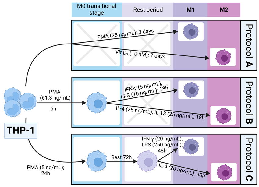
DOI:10.3390/biomedicines11020608
2. UA BIOSCIENCE New Product Recommendation
UA BIOSCIENCE has launched the new Human THP-1 Cell Polarization Induction Kit (Catalog No. UA090040), providing a one-stop, highly stable solution for constructing in vitro models for macrophage functional research. This kit includes rigorously validated cytokine combinations and detailed protocols, enabling researchers to easily achieve directional polarization of THP-1 monocytes into M1 or M2-type macrophages.
Why Choose UA BIOSCIENCE's THP-1 Polarization Solution?
✅ High Efficiency and Stability
Optimized specifically for THP-1 cells, with high M1/M2 polarization efficiency and excellent batch-to-batch reproducibility.
✅ Easy Operation
Standardized protocols reduce experimental optimization time, enabling rapid construction of models for inflammation, cancer, metabolism, and other disease research.
✅ Broad Applications
Suitable for immune regulation mechanisms, anti-inflammatory drug screening, tumor microenvironment studies, and more.
3. Experimental Protocol
Human THP-1 Cell Polarization Induction Kit (Catalog No. UA090040) Protocol:
Pre-Polarization Preparation:
Cells used for polarization should not be of excessively high passage number. Before induction, detect the expression of CD14 and CD11b in THP-1 cells using flow cytometry. Only THP-1 cells with no or low expression of these markers should be used for subsequent experiments.
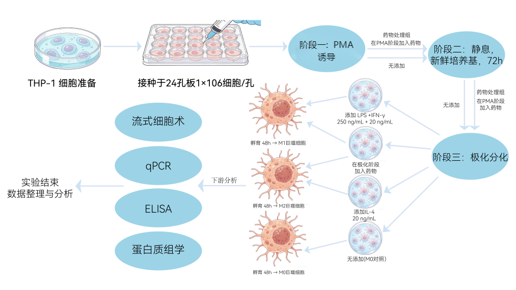
M1/M2 Induction
1. M1 Induction:
Use M0 induction medium (1640 complete medium containing 100 ng/mL PMA) to seed THP-1 cells (in good condition, viability >95%, <20 passages) at a density of 5×10⁵ cells/mL in a 10 cm dish (10 mL), and culture for 24 h to construct M0 macrophages (adherent cells with pseudopod extension). Gently wash 1-2 times with pre-warmed PBS to remove PMA. Replace with M1 induction medium (1640 complete medium containing 100 ng/mL LPS and 20 ng/mL IFN-γ) and culture for 48 h.
Negative control: Use M0 induction medium (1640 complete medium containing 100 ng/mL PMA) to seed THP-1 cells (in good condition, viability >95%, <20 passages) at a density of 5×10⁵ cells/mL in a 10 cm dish (10 mL), and culture for 24 h to construct M0 macrophages (adherent cells with pseudopod extension). Gently wash 1-2 times with pre-warmed PBS to remove PMA. Replace with complete medium and culture for 48 h. It is recommended to harvest samples using Accutase for analysis.
2. M2 Induction:
Use M0 induction medium (1640 complete medium containing 100 ng/mL PMA) to seed THP-1 cells (in good condition, viability >95%, <20 passages) at a density of 5×10⁵ cells/mL in a 10 cm dish (10 mL), and culture for 24 h to construct M0 macrophages (adherent cells with pseudopod extension). Gently wash 1-2 times with pre-warmed PBS to remove PMA, replace with complete medium, and rest for 24 h. Then replace with M2 induction medium (1640 complete medium containing 20 ng/mL IL-4 and 20 ng/mL IL-13) and culture for 24 h.
Negative control: Use M0 induction medium (1640 complete medium containing 100 ng/mL PMA) to seed THP-1 cells (in good condition, viability >95%, <20 passages) at a density of 5×10⁵ cells/mL in a 10 cm dish (10 mL), and culture for 24 h to construct M0 macrophages (adherent cells with pseudopod extension). Gently wash 1-2 times with pre-warmed PBS to remove PMA, replace with complete medium, and rest for 24 h. It is recommended to harvest samples using Accutase for analysis.
4. Application Scenarios
- Inflammation and Autoimmune Diseases: Simulate M1-mediated inflammatory responses for anti-inflammatory drug screening and inflammasome mechanism research.
- Cancer Immunology: Construct M2-type tumor-associated macrophage models to study immune escape mechanisms and repolarization drugs.
- Infection Immunology: Establish pathogen-macrophage interaction models to evaluate killing functions and immune escape strategies.
- Drug Screening: Use M1/M2 markers as indicators to quantitatively assess the immunomodulatory activity of candidate compounds.
- Tissue Repair: Simulate M2-mediated tissue remodeling processes to evaluate pro-healing materials and fibrosis mechanism research.
This article is reviewed and published by the technical expert team of UA
Disclaimer: This article partially utilizes artificial intelligence assistance in its creation. If any content involves copyright or intellectual property issues, please let us know and we promise to verify and remove it as soon as possible.
Purchase recombinant protein, choose 
UA protein focuses on providing various protein reagents, raw materials, and services required for drug research and development, cell therapy, gene therapy, and basic scientific research, including drug target proteins, immune checkpoint proteins, cytokines, tool enzymes, customized protein expression, and full-length transmembrane protein development. Nanjing Univ is committed to providing customers with high-quality products and professional services, and building a High-tech Biological Enterprise with International Competitiveness.







 Business license (three certificates in one)

