




Technical Features and Applications of the UA-Glo® Nano-luc Live Cell Detection System


Real-time, quantitative monitoring of molecular events in living cells, such as protein-protein interactions, protein stability, and gene expression regulation, is a core requirement in cell biology research and drug discovery.
- Recent Advances
- Product Information
I. Introduction
Real-time, quantitative monitoring of molecular events in living cells, such as protein-protein interactions, protein stability, and gene expression regulation, is a core requirement in cell biology research and drug discovery. Reporter gene technologies based on luciferase are widely used due to their high sensitivity, broad linear range, and operational simplicity. In recent years, the emergence of novel small-molecule luciferases (NanoLuc) and their fragmentation techniques has further enhanced detection performance. This article introduces a live-cell detection system based on split NanoLuc technology, detailing its technical principles, core advantages, and standard operating procedures.
II. Technical Principles and Design Basis
This system is constructed based on split luciferase complementation technology. Its core technical component involves dividing the micro-luciferase (NanoLuc) into two peptide fragments with no independent catalytic activity at specific structural sites: a large fragment (Large Bit, LgBit) and a small fragment (Small Bit, SmBit).
In application design, these two fragments can be fused with the target proteins under study. When the target proteins interact or are part of the same complex, LgBit and SmBit come into close spatial proximity, reconstructing the complete NanoLuc active conformation. In the presence of cell-permeable, high-sensitivity substrates, the reconstructed enzyme catalyzes the substrate to produce high-intensity, sustainable bioluminescence signals.
Depending on the inherent affinity between the fragments, this technology can be adapted to different research scenarios. High-affinity fragment combinations can spontaneously and rapidly complement, making them suitable for monitoring total protein expression levels or serving as the basis for biosensors. In contrast, low-affinity fragment complementation driven by target protein interactions is specifically designed to study the dynamic formation of protein complexes.
III. Core Advantages of the Live-Cell Detection System
Combining the above technology with optimized live-cell detection reagents forms a complete system with multiple significant technical advantages, particularly suitable for high-throughput screening (HTS) and real-time kinetic analysis.
1. High Sensitivity and Low Background: NanoLuc enzyme itself exhibits higher specific activity than traditional firefly or Renilla luciferases. Combined with optimized cell-permeable substrates, it can detect weak interaction signals in living cells. Since uncomplemented fragments are inactive, background signals are extremely low, resulting in a high signal-to-noise ratio.
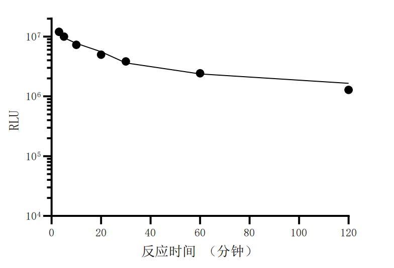
2. Stable Signals, Suitable for HTS: Under detection conditions, the luminescence signal generated by the reconstructed enzyme has a half-life of approximately 2 hours. This "glow-type" signal characteristic allows for batch processing of samples without strict timing, greatly facilitating the operation of automated high-throughput screening platforms.
3. Homogeneous Detection, Maintaining Cell Viability: The system is equipped with cell-permeable substrates, eliminating the need for cell lysis. Simply adding the detection reagent directly to the culture plate enables real-time reading of events within live cell populations, supporting dynamic monitoring at multiple time points or subsequent functional analysis of cells.
4. Flexible Operation and Adaptability: The product components are optimized to allow selection of different detection methods based on experimental objectives (e.g., pursuing the highest signal, optimal cell state, or simplest operational steps), and are compatible with microplates of various specifications.
IV. Key Points of Standardized Operating Procedures
To ensure the accuracy and reproducibility of detection results, standardized operating procedures must be followed, with key steps including:
1. Cell Model Construction: Fuse the target genes with the coding frames of LgBit and SmBit fragments, respectively, to construct expression vectors. Introduce these into suitable host cells (e.g., HEK293) via transient transfection or stable cell line establishment.
2. Cell Seeding and Treatment: Seed an appropriate number of cells in white, clear-bottom cell culture plates to ensure optimal cell density during experiments. Perform drug treatments, genetic interventions, or other operations as per the experimental design, and culture until the target proteins are expressed and interactions occur at the desired time points.
3. Reagent Equilibrium and Preparation: Equilibrate the detection buffer to room temperature (22-25°C). Thaw the NanoLuc substrate on ice and freshly prepare a 1× detection working solution by diluting it 1:200 with the buffer, ensuring light protection.
4. Detection and Signal Reading: Equilibrate the cell culture plate to room temperature. Optionally, remove the culture medium and add the detection solution directly (for the highest signal) or retain some medium to maintain better cell conditions. After adding the detection solution, incubate at room temperature in the dark for 10 minutes to allow the reaction to equilibrate. Finally, use a multifunctional microplate reader equipped with a luminescence detection module to read the luminescence signal values.
5. Data Interpretation: The intensity of the luminescence signal directly reflects the degree of complementation between LgBit and SmBit, thereby quantitatively characterizing the interaction strength between target proteins or the expression level of target proteins.
V. Conclusion
In summary, the UA-Glo® live-cell detection system, based on split NanoLuc technology, achieves high-sensitivity, real-time, homogeneous detection of molecular events in living cells through innovative enzyme fragment design and optimized cell-permeable substrates. Its stable signals, simple operation, and low background make it a powerful tool for exploring protein functions in basic research and conducting high-throughput compound screening in drug development. Strict adherence to standardized operating procedures is key to leveraging the system's performance and obtaining reliable data.
VI. Which Manufacturers Provide the UA-Glo® Nano-luc Live-Cell Detection System?
Nanjing UA-Bio Technology Co., Ltd. (UA-Bio) has independently developed the "UA-Glo® Nano luciferase Live Cell Assay System", a high-performance bioluminescence detection system specifically designed for real-time dynamic analysis of living cells. This system employs optimized NanoLuc technology to accurately and efficiently monitor the dynamic expression of reporter genes in live cells, providing stable and reliable standardized solutions for real-time cell analysis, drug time-effect studies, and signal pathway tracking.
| Core Product Advantages |
|---|
| Ultra-High Brightness and Real-Time Monitoring Capability: This system uses engineered NanoLuc luciferase to produce luminescence signals far exceeding those of traditional firefly luciferase. Its exceptional brightness and optimized substrate permeability enable real-time, continuous tracking of gene expression dynamics in live cells without cell lysis, presenting complete time-effect curves. |
| Excellent Cell Compatibility: The optimized substrate formulation exhibits minimal cytotoxicity, preserving normal cell growth and physiological functions, and supports long-term, multi-timepoint continuous monitoring. This non-destructive detection mode allows sustained tracking of cellular responses within the same culture system, significantly enhancing experimental efficiency and data quality. |
| Outstanding Batch-to-Batch Consistency and Stability: Leveraging an internationally leading luminescence detection technology platform and standardized production processes, combined with a stringent quality control system, ensures high purity, excellent long-term stability, and superior batch-to-batch consistency for all system components. This provides a solid foundation for long-term, continuous live-cell research. |
| Homogeneous, Wash-Free, Flexible, and Efficient Experimental Mode: This system adopts a simple "add-and-read" homogeneous operation mode, requiring only the addition of a single detection reagent to live cell culture wells, with no need for cumbersome separation or washing steps. Its optimized formulation is compatible with multi-well plate (96/384-well) automation platforms, making it adaptable for various applications such as high-throughput drug screening, GPCR signal dynamic analysis, circadian rhythm studies, and cell proliferation monitoring. |
| Stable and Persistent Glow-Type Signals: The kit is specially formulated to produce uniform, stable "glow-type" luminescence signals with prolonged duration, providing ample time windows for long-term dynamic monitoring and ensuring data reliability and reproducibility. |
| Complete Solutions and Professional Support: We provide fully validated standard experimental protocols, typical dynamic curves, and detailed result interpretation guidelines to help you quickly establish stable and reproducible experimental workflows. Nanjing UA-Bio's professional technical team offers comprehensive support for research design, experimental optimization, and data analysis. |
Nanjing UA-Bio Technology Co., Ltd. is dedicated to providing cutting-edge, high-quality core reagents and tools for immunology, cell therapy, and innovative drug development. For detailed technical parameters, validation data, or specific application inquiries regarding the "UA-Glo® Nano luciferase Live Cell Assay System" (Catalog No.: UA070110), please feel free to contact us.
This article is reviewed and published by the technical expert team of UA
Disclaimer: This article partially utilizes artificial intelligence assistance in its creation. If any content involves copyright or intellectual property issues, please let us know and we promise to verify and remove it as soon as possible.
Purchase recombinant protein, choose Nanjing UA-Bio
UA protein focuses on providing various protein reagents, raw materials, and services required for drug research and development, cell therapy, gene therapy, and basic scientific research, including drug target proteins, immune checkpoint proteins, cytokines, tool enzymes, customized protein expression, and full-length transmembrane protein development. Youai is committed to providing customers with high-quality products and professional services, and building a High-tech Biological Enterprise with International Competitiveness.







 Business license (three certificates in one)