




Macrophage Polarization and Its Role in the Tumor Microenvironment


Macrophages are highly heterogeneous immune cells with significant ontogenic differences in their origins. During embryonic development, certain tissue-resident macrophages, such as Langerhans cells in the skin, microglia in the central nervous system, Kupffer cells in the liver, and alveolar macrophages, originate from yolk sac and fetal liver precursors. These cells maintain tissue homeostasis through self-renewal.
- Recent Advances
- Product Information
I. Origin and Tissue Distribution of Macrophages
Macrophages are highly heterogeneous immune cells with significant developmental differences in their origin. During embryonic development, some tissue-resident macrophages originate from yolk sac and fetal liver precursors, such as skin Langerhans cells, central nervous system microglia, liver Kupffer cells, and alveolar macrophages, which maintain tissue homeostasis through self-renewal. Under inflammatory or pathological conditions, bone marrow-derived monocytes are recruited to specific tissues, differentiating into macrophages and gradually acquiring phenotypic characteristics similar to embryonic-derived macrophages. Different tissue microenvironments endow macrophages with unique transcriptional profiles and epigenetic features. In the tumor microenvironment (TME), the origin of macrophages is more complex, involving both the proliferation of tissue-resident macrophages and the infiltration and differentiation of bone marrow-derived monocytes. In basic research, the mouse RAW264.7 cell polarization kit can be used to simulate different polarization states of macrophages, providing an important tool for investigating their functional regulation mechanisms.
II. Functional Plasticity and Polarization Regulation of Macrophages
Macrophages exhibit remarkable functional plasticity and can undergo phenotypic switching in response to different microenvironmental signals. Classical M1 polarization is induced by interferon-γ (IFN-γ) and bacterial products such as lipopolysaccharide (LPS), resulting in a pro-inflammatory phenotype characterized by high expression of inducible nitric oxide synthase (iNOS) and tumor necrosis factor-α (TNF-α), which participate in pathogen clearance and tumor cell killing. M2 polarization is induced by interleukin-4 (IL-4) and interleukin-13 (IL-13), resulting in an anti-inflammatory phenotype characterized by high expression of arginase 1 (Arg1) and interleukin-10 (IL-10), which participate in tissue repair and immune regulation. However, in vivo, macrophages often exhibit mixed phenotypes, and the simple M1/M2 dichotomy cannot fully capture their functional diversity. In the tumor microenvironment, multiple cytokines, such as colony-stimulating factor 1 (CSF-1), chemokine ligand 2 (CCL2), and IL-10, collectively regulate the functional phenotype of tumor-associated macrophages (TAM). The mouse RAW264.7 cell polarization kit can simulate the M1 and M2 polarization processes of macrophages in vitro, providing an experimental basis for analyzing their phenotypic switching mechanisms.
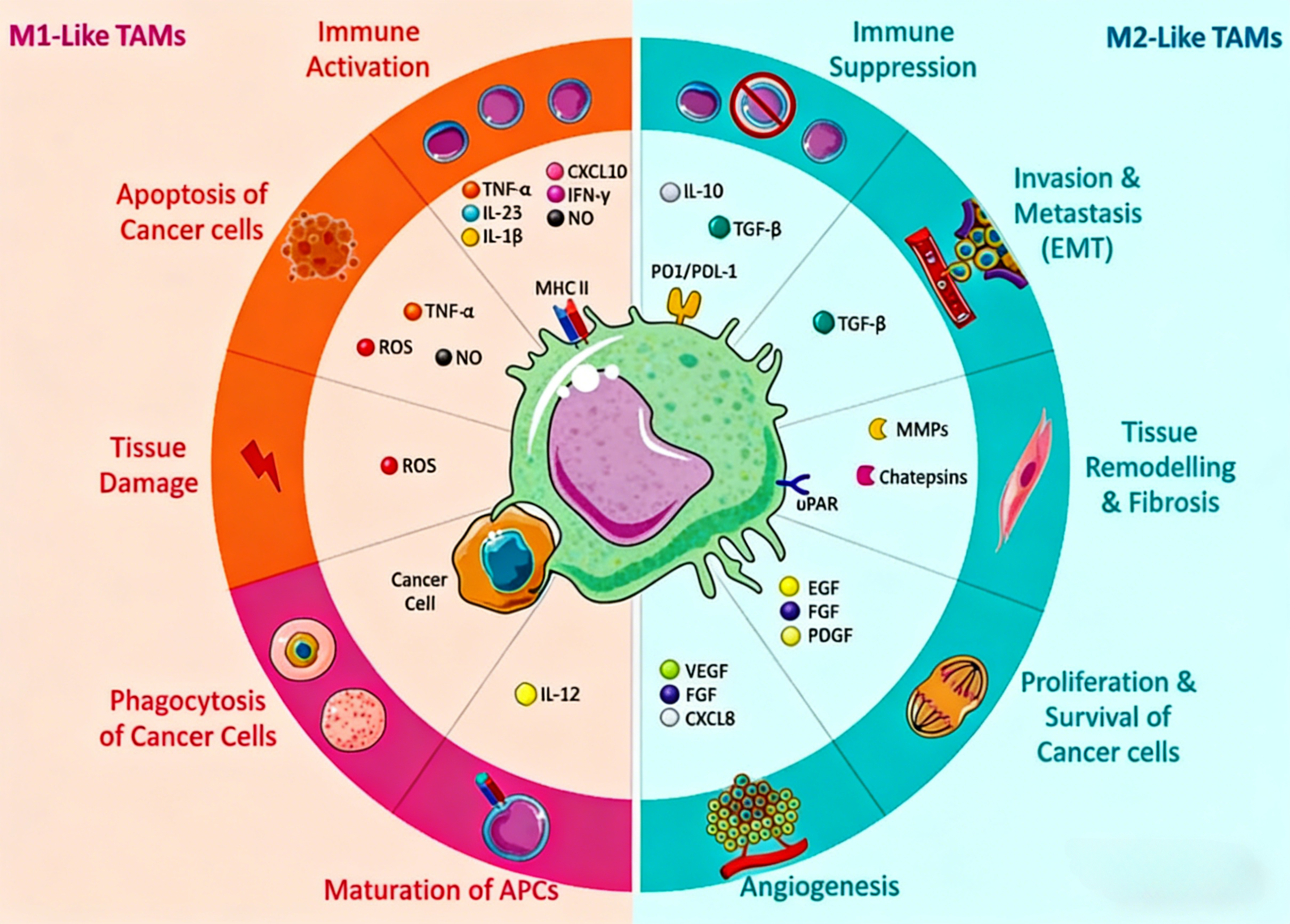
III. Regulatory Mechanisms and Functional Heterogeneity of Tumor-Associated Macrophages
Tumor-associated macrophages typically exhibit a pro-tumor phenotype in the tumor microenvironment, and their functional regulation involves multi-layered molecular mechanisms. Various factors secreted by tumor cells and stromal cells drive macrophages toward an immunosuppressive phenotype. Single-cell sequencing studies have revealed the high heterogeneity of TAM, with different subpopulations displaying unique transcriptional features and functional attributes, such as pro-angiogenic subpopulations, immunosuppressive subpopulations, and tissue-remodeling subpopulations. The expression of inhibitory receptors on macrophages, such as programmed death receptor 1 (PD-1), can negatively regulate their phagocytic function toward tumor cells, affecting anti-tumor immune responses. Additionally, metabolic factors in the tumor microenvironment, such as hypoxia and lactate accumulation, also participate in regulating macrophage functional polarization. The cell models constructed using the mouse RAW264.7 cell polarization kit can be used to study the influence of tumor microenvironmental factors on macrophage polarization, providing a theoretical basis for targeted therapies.
IV. Role of Macrophages in Tumor Progression and Metastasis
Tumor-associated macrophages participate in tumor progression through multiple mechanisms. In primary tumor growth, macrophages promote tumor angiogenesis by secreting vascular endothelial growth factor (VEGF), providing nutrients for tumor growth. Simultaneously, macrophages secrete matrix metalloproteinases (MMPs) that participate in extracellular matrix remodeling, facilitating tumor cell invasion. Macrophages play important roles in multiple steps of tumor metastasis: they promote angiogenesis to create conditions for hematogenous dissemination of tumor cells; they are recruited to distant organs during the formation of pre-metastatic niches, creating a microenvironment suitable for tumor cell colonization; and in lymphatic metastasis, specific macrophage subpopulations participate in tumor cell diffusion by regulating lymphangiogenesis. Macrophages in different tissue locations have organ-specific effects on tumor metastasis. The mouse RAW264.7 cell polarization kit can be used to investigate the functional differences of macrophages in different polarization states during tumor metastasis.
V. Tumor Treatment Strategies Targeting Macrophages
Macrophages have become important targets for tumor therapy. Chemotherapeutic drugs such as doxorubicin can induce immunogenic cell death (ICD), activating macrophage-mediated anti-tumor immunity. Certain chemotherapeutic drugs, such as gemcitabine, can reverse macrophage polarization, switching them from a pro-tumor phenotype to an anti-tumor phenotype, thereby enhancing therapeutic efficacy. The effects of radiotherapy are regulated by the gut microbiome, with macrophages acting as a bridge. Anti-angiogenic therapy can induce tumor vascular normalization and reshape macrophage phenotypes, but myeloid cells can also mediate resistance through compensatory pathways. The efficacy of immune checkpoint blockade therapy is closely related to the functional state of TAM, and the expression of immunosuppressive molecules such as PD-L1 by macrophages contributes to treatment resistance. In specific locations such as liver metastases, tissue-resident macrophages negatively regulate immunotherapy efficacy by clearing T cells. Macrophage-targeted treatment strategies, such as those targeting CSF-1 receptor, are being explored in clinical trials. The mouse RAW264.7 cell polarization kit provides an important in vitro research model for screening anti-tumor drugs that target macrophage polarization states.
VI. Which Manufacturers Provide Mouse RAW264.7 Cell Polarization Kits?
Nanjing UA-Bio Technology Co., Ltd. (UA-Bio) has independently developed the "Mouse RAW264.7 Cell Polarization Induction Kit II", a high-performance cell culture system specifically designed to induce polarization in the mouse RAW264.7 macrophage cell line. This kit uses an optimized combination of induction factors to efficiently and stably drive RAW264.7 cells toward classical activation (M1) or alternative activation (M2) macrophage polarization, providing standardized and reliable solutions for research in inflammation mechanisms, immune regulation evaluation, drug screening, and other fields.
| Core Advantages of the Product |
|---|
| Efficient Polarization and Directional Induction: The kit employs a carefully balanced combination of induction factors (including LPS, IFN-γ, IL-4, IL-13, etc.) to simulate microenvironmental signals for macrophage polarization in vivo, enabling efficient induction of RAW264.7 cells toward M1 or M2 polarization. The optimized induction system promotes polarization while effectively maintaining the expression of subtype-specific markers and functional characteristics, including M1-type iNOS, TNF-α secretion capacity, and M2-type Arg1 and IL-10 expression levels. |
| Excellent Batch-to-Batch Consistency and Stability: Relying on an internationally leading cell culture reagent development platform and standardized production processes, combined with a strict quality control system, each batch of reagents exhibits stable polarization efficiency, consistent cell function, and excellent long-term stability. This provides solid and reliable quality assurance for long-term and continuous macrophage functional research. |
| Flexible and Convenient Operating System: The kit provides a complete polarization protocol and optimized culture system, making the operation simple and efficient. Its formulation is compatible with various culture vessels and detection platforms, flexibly applicable to RAW264.7 cell M1/M2 polarization induction, inflammatory factor secretion analysis, phagocytic function evaluation, and signaling pathway research, among other application needs. |
| Comprehensive Solutions and Professional Support: We provide fully validated standard experimental protocols, typical polarization efficiency data, and detailed result interpretation guidelines to help you quickly establish stable and reproducible RAW264.7 cell polarization processes. The Nanjing UA-Bio professional technical team offers comprehensive technical consultation and support for your research design, experimental optimization, and data analysis. |
Nanjing UA-Bio Technology Co., Ltd. is committed to providing cutting-edge, high-quality core reagents and tools for immunology, cell therapy, and innovative drug development. For detailed technical parameters, validation data, or specific application inquiries regarding the "Mouse RAW264.7 Cell Polarization Induction Kit II" (Catalog No.: UA090038), please feel free to contact us.
This article is reviewed and published by the technical expert team of UA
Disclaimer: This article partially utilizes artificial intelligence assistance in its creation. If any content involves copyright or intellectual property issues, please let us know and we promise to verify and remove it as soon as possible.
Purchase recombinant protein, choose Nanjing UA-Bio
UA protein focuses on providing various protein reagents, raw materials, and services required for drug research and development, cell therapy, gene therapy, and basic scientific research, including drug target proteins, immune checkpoint proteins, cytokines, tool enzymes, customized protein expression, and full-length transmembrane protein development. Youai is committed to providing customers with high-quality products and professional services, and building a High-tech Biological Enterprise with International Competitiveness.







 Business license (three certificates in one)