




Application of Mouse Normal Small Intestinal Organoid Cytokine Panel in the Study of Crohn's Disease Mechanisms


Small intestinal organoids are miniature tissue structures formed through the self-organization of adult stem cells from the small intestine under three-dimensional culture conditions. They can highly replicate the physiological structure and cellular composition of the intestinal epithelium, containing various cell types such as intestinal stem cells, Paneth cells, goblet cells, enteroendocrine cells, and absorptive enterocytes.
- Recent Advances
- Product Information
I. The Value of Small Intestinal Organoids in Intestinal Disease Research
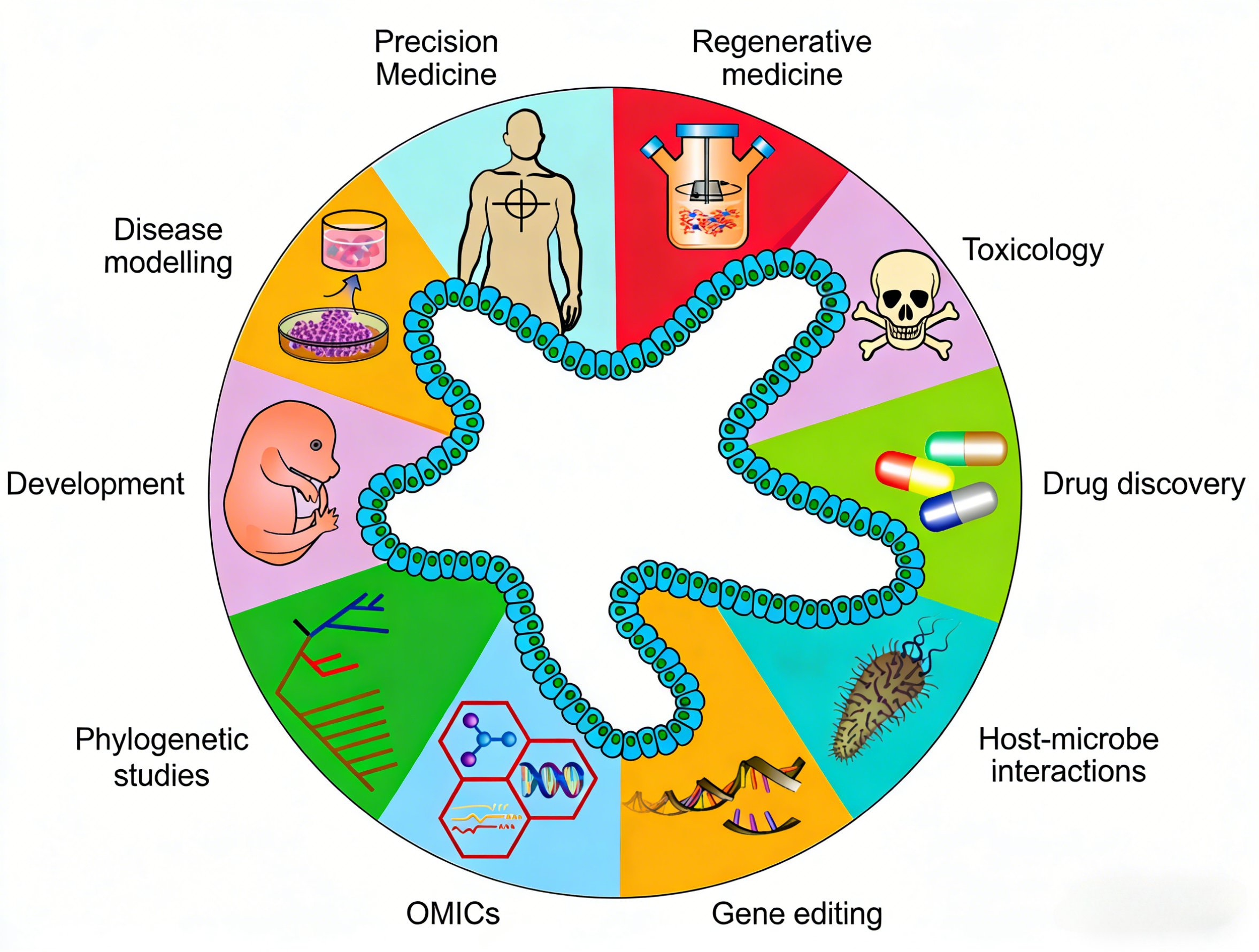
Small intestinal organoids are miniature tissue structures self-organized from small intestinal adult stem cells under three-dimensional culture conditions, capable of highly replicating the physiological structure and cellular composition of the intestinal epithelium, including intestinal stem cells, Paneth cells, goblet cells, enteroendocrine cells, and absorptive enterocytes, among others. This culture system mimics the in vivo stem cell microenvironment by adding specific cytokine combinations, maintaining the self-renewal and multi-directional differentiation potential of stem cells. Small intestinal organoid models hold significant value in the study of intestinal infections and the mechanisms of inflammatory bowel disease (IBD), and also provide an ideal in vitro research platform for analyzing host-microbe interactions in the gut. In basic research, using the mouse normal small intestinal organoid cytokine kit can simulate the growth microenvironment of small intestinal organoids, offering an important tool for studying the intestinal epithelium's response mechanisms to pathogens.
II. The Potential Association Between Crohn's Disease and Mycobacterium avium
Crohn's disease (CD), as a chronic recurrent inflammatory bowel disease, has always been a research focus in the field of intestinal diseases. Mycobacterium avium subspecies paratuberculosis (MAP) is considered a potential trigger, but the mechanisms linking the two and the early response patterns of the intestinal epithelium remain unclear. To explore the potential association between MAP infection and Crohn's disease, the research team constructed a MAP-infected mouse small intestinal organoid model. Transcriptome analysis and comparison with Crohn's disease patient-derived mouse models revealed a large number of overlapping differentially expressed genes and enriched pathways, confirming that infection can induce intestinal epithelium to produce inflammatory features highly similar to Crohn's disease. The study identified Mmp7 as a key molecular marker linking MAP infection to Crohn's disease-like pathology, with its mediated epithelial remodeling and inflammatory responses significantly activated in both models. Additionally, the TLR2 signaling pathway was found to regulate the directional recruitment of macrophages to infected organoids, providing new research insights and potential therapeutic targets for Crohn's disease.
III. Establishment of the Mouse Small Intestinal Organoid Infection Model
To investigate the potential association between MAP infection and Crohn's disease, the research team pioneered the construction of a mouse small intestinal organoid infection model. Successfully cultured mouse small intestinal organoids could stably expand and maintain in vitro. After co-culturing with fluorescently labeled MAP, live-cell confocal imaging showed that bacterial load within the organoids significantly increased over time; quantitative analysis further confirmed a progressive increase in MAP numbers. When MAP was directly microinjected into the Matrigel between two organoids, directional migration of basolateral cells toward the bacterial location was observed, indicating that epithelial cells possess the ability to sense and respond to bacterial stimuli. Cytoskeleton staining results verified that MAP could successfully invade and localize within mouse small intestinal organoids, establishing a reliable in vitro infection model for subsequent mechanistic studies. During this process, the mouse normal small intestinal organoid cytokine kit provided standardized conditions for the stable culture and expansion of organoids, supporting the construction and maintenance of the infection model.
IV. Cellular Response Mechanisms Induced by Mycobacterium avium Infection
MAP can actively invade intestinal epithelial cells and induce directional cellular migration. The study found that MAP can invade intestinal epithelial cells from the basolateral side and establish intracellular colonization, while also inducing directional migration of epithelial cells toward the bacterial location, revealing the active response capability of the intestinal epithelium to pathogens. This phenomenon suggests that intestinal epithelial cells are not just a physical barrier but can actively sense bacterial stimuli and respond directionally. Using the mouse small intestinal organoid infection model, the spatiotemporal dynamics of epithelial cell migration toward infection sites can be observed and recorded in real-time, providing a unique perspective for studying host-pathogen interactions. Organoids cultured with the mouse normal small intestinal organoid cytokine kit exhibit excellent physiological activity and response capability, making them an ideal tool for studying such cellular behaviors.
V. The Role of the TLR2 Signaling Pathway in Macrophage Recruitment
The study clarified that the TLR2 signaling pathway regulates the directional recruitment of macrophages to infected organoids. Macrophages can directionally migrate toward MAP-infected organoids, form clusters, and eventually infiltrate the organoids, a process regulated by the TLR2 signaling pathway, simulating early immune events of granuloma formation. This discovery reveals the molecular mechanism by which the innate immune system recognizes MAP infection and the interactions between immune cells and epithelial cells. In co-culture systems, observing the directional migration and infiltration of immune cells when macrophages are co-cultured with infected organoids provides a controllable in vitro model for studying early intestinal inflammatory events. The organoid culture system supported by the mouse normal small intestinal organoid cytokine kit can be used to establish such immune-epithelial co-culture models, exploring the molecular mechanisms of immune cell recruitment.
VI. Discovery of Mmp7 as a Key Molecular Marker
The study identified Mmp7 as a key molecular marker linking MAP infection to Crohn's disease-like inflammation. MAP infection can activate core inflammatory pathways such as TNF-α/NF-κB and IL-6/JAK/STAT3, with IBD signaling pathways significantly enriched in both infected organoids and Crohn's disease mouse models. Mmp7 was significantly upregulated in both models, with its mediated epithelial remodeling and inflammatory responses being core features shared by both, providing potential therapeutic targets for Crohn's disease. Transcriptome analysis revealed a large number of overlapping differentially expressed genes and enriched pathways between infected organoids and Crohn's disease models, confirming that infection can induce intestinal epithelium to produce inflammatory features highly similar to Crohn's disease. This discovery not only reveals the molecular link between infection and disease but also provides a theoretical basis for developing new therapeutic strategies.
VII. Which Manufacturers Provide the Mouse Normal Small Intestinal Organoid Cytokine Kit?
Nanjing UA-Bio Biotechnology Co., Ltd. (UA-Bio) has independently developed the "Small Intestinal Organoid Cytokine Set, Mouse", a high-performance cytokine kit specifically designed for constructing and maintaining mouse normal small intestinal organoids. This kit employs an optimized cytokine combination to simulate the microenvironment signals for small intestinal crypt stem cell proliferation and differentiation, supporting the long-term expansion and organoid formation of mouse small intestinal stem/progenitor cells. It provides standardized and reliable solutions for research in intestinal development, nutrient absorption mechanisms, and drug evaluation.
| Core Product Advantages |
|---|
| Optimized Organoid Growth Support: The kit uses carefully selected and proportioned cytokine combinations, including key signaling pathway regulators (such as EGF, Noggin, R-spondin, Wnt agonists, etc.), to precisely simulate the microenvironment signals for small intestinal stem cell proliferation and differentiation. It efficiently supports the establishment, long-term expansion, and intestinal epithelial lineage differentiation of mouse normal small intestinal organoids. The optimized cytokine ratio promotes organoid formation while effectively maintaining the cellular diversity and functional integrity of the intestinal villus-crypt structure. |
| Excellent Batch-to-Batch Consistency and Stability: Relying on an internationally leading cytokine development platform and standardized production processes, combined with a strict quality control system, each batch of reagents ensures stable biological activity, consistent organoid support capability, and excellent long-term stability. It provides solid and reliable quality assurance for long-term and continuous small intestinal organoid research. |
| Flexible and Convenient Operating System: The kit provides a complete organoid culture protocol and optimized cytokine combinations, making operations simple and fast. Its formulation system is compatible with various organoid culture matrices and containers, flexibly applicable to multiple application needs such as the establishment, expansion, differentiation research, cryopreservation, and drug screening of mouse normal small intestinal organoids. |
| Complete Solutions and Professional Support: We provide fully validated standard experimental protocols, typical organoid growth data, and detailed result interpretation guides to help you quickly establish a stable and reproducible mouse small intestinal organoid culture system. Nanjing UA-Bio's professional technical team offers comprehensive and professional technical consultation and support for your research design, experimental optimization, and data analysis. |
Nanjing UA-Bio Biotechnology Co., Ltd. is always committed to providing cutting-edge, high-quality core reagents and tools for immunology, cell therapy, and innovative drug development. For detailed technical parameters, validation data, or specific application inquiries regarding the "Small Intestinal Organoid Cytokine Set, Mouse" (Catalog No.: UA090043), please feel free to contact us.
This article is reviewed and published by the technical expert team of UA
Disclaimer: This article partially utilizes artificial intelligence assistance in its creation. If any content involves copyright or intellectual property issues, please let us know and we promise to verify and remove it as soon as possible.
Purchase recombinant protein, choose Nanjing UA-Bio
UA protein focuses on providing various protein reagents, raw materials, and services required for drug research and development, cell therapy, gene therapy, and basic scientific research, including drug target proteins, immune checkpoint proteins, cytokines, tool enzymes, customized protein expression, and full-length transmembrane protein development. Youai is committed to providing customers with high-quality products and professional services, and building a High-tech Biological Enterprise with International Competitiveness.







 Business license (three certificates in one)