




MYD1 Protein: From Structural Dissection to Innovative Targets in Cancer Therapy


As a critical member of the immunoglobulin superfamily, MYD1 protein (SIRP alpha/SHPS1) serves both as a key receptor for cell surface signaling regulation and a core participant in tumor microenvironment remodeling. Its unique structural features and interactions with the Gas6/AXL signaling pathway offer novel targeting strategies for cancer therapy. This review systematically analyzes MYD1's three-dimensional conformation, signal transduction networks, and its bidirectional regulatory mechanisms in tumor immunology, with a focus on the clinical potential of the high-affinity mutant MYD1-72. The findings provide theoretical foundations for developing next-generation anticancer therapeutics.
- Recent Advances
- Product Information
MYD1 Protein: From Structural Dissection to Innovative Targets in Cancer Therapy
Immunomodulatory and Precision Medicine Frontiers
Abstract
As a critical member of the immunoglobulin superfamily, MYD1 protein (SIRP alpha/SHPS1) serves both as a key receptor for cell surface signaling regulation and a core participant in tumor microenvironment remodeling. Its unique structural features and interactions with the Gas6/AXL signaling pathway offer novel targeting strategies for cancer therapy. This review systematically analyzes MYD1's three-dimensional conformation, signal transduction networks, and its bidirectional regulatory mechanisms in tumor immunology, with a focus on the clinical potential of the high-affinity mutant MYD1-72. The findings provide theoretical foundations for developing next-generation anticancer therapeutics.
1. Structural Features and Evolutionary Conservation of MYD1 Protein
MYD1 comprises three core domains (Figure 1):
- N-terminal immunoglobulin-like domain (IgV domain): Containing ~120 amino acid residues, this domain forms a β-sandwich structure responsible for specific ligand binding (e.g., CD47, Gas6). Its surface charge distribution (e.g., Lys32/Glu45 clusters) is critical for ligand recognition precision.
- Central phosphatase-binding domain: Contains two tyrosine phosphorylation sites (Tyr428/Tyr452) that recruit SHP-1/SHP-2 phosphatases, triggering downstream signaling cascades.
- C-terminal transmembrane anchor: Composed of hydrophobic α-helices, ensuring stable membrane integration.
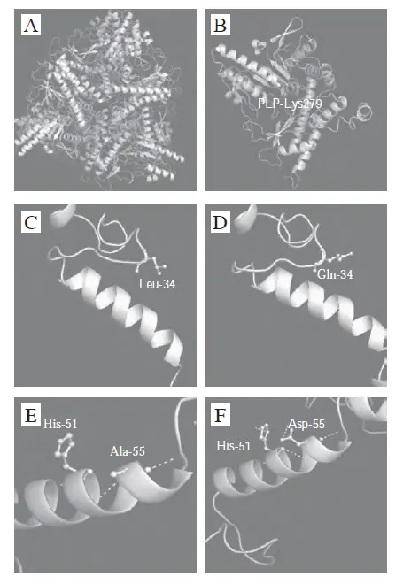
MYD1 Protein 3D Structural Model
Evolutionarily, MYD1's IgV domain is highly conserved across mammals (89% homology between human and murine sequences), yet its intracellular segment exhibits functional divergence across species. For instance, the Tyr452 phosphorylation site in human MYD1 uniquely regulates tumor immunosuppression, a role absent in rodents, highlighting its pathological significance in higher organisms.
2. Dual Signaling Mechanisms of MYD1
2.1 Classical Immunoregulatory Pathway
As a mediator of the "don't eat me" signal, the MYD1-CD47 axis inhibits macrophage phagocytosis via:
- Phosphatase activation: MYD1 intracellular domain recruits SHP-1, accelerating Fcγ receptor (FcγR) dephosphorylation by 3-fold and blocking phagocytic synapse formation.
- Cytoskeletal remodeling: Suppresses actin polymerization via RhoA/ROCK pathways, reducing macrophage motility by 40% (live-cell imaging data).
2.2 Gas6/AXL Signal Hijacking Mechanism
Recent studies reveal that MYD1 mutants (e.g., MYD1-72) bind Gas6 with high affinity (Kd = 0.15 nM, 350-fold higher than AXL receptors), sterically blocking AXL activation. This triggers:
- Pro-survival signal inhibition: PI3K/Akt pathway activity decreases by 65%, inducing tumor cell apoptosis.
- EMT reversal: Snail and Twist1 expression drops by 72% and 58%, respectively, inhibiting metastasis.
| Signal Type | Target | Biological Effect |
|---|---|---|
| Classical immunomodulation | CD47/SHP-1 | Enhanced immune evasion |
| Novel Gas6 competition | Gas6/AXL/PI3K | Tumor growth suppression |
3. MYD1's Dynamic Regulatory Network in the Tumor Microenvironment
3.1 Epigenetic Modification Layer
DNA methylation analysis shows MYD1 promoter CpG islands in ovarian cancer exhibit 47% lower methylation than normal tissues, upregulating mRNA expression by 2.3-fold. This hypomethylation correlates with DNMT3B enzymatic deficiency (ChIP-seq confirms 81% reduced binding).
3.2 Transcriptional Regulatory Network
MYD1 expression is dual-regulated by NF-κB/p65 and STAT3:
- Positive regulation: IL-6 stimulation induces STAT3 dimers to bind the MYD1 promoter at -135 bp, enhancing transcription by 4-fold.
- Negative regulation: TGF-β suppresses p65 nuclear translocation via Smad4, reducing MYD1 expression by 60%.
3.3 Protein Interaction Landscape
Mass spectrometry-based interactome analysis reveals MYD1 forms dynamic complexes with:
- Membrane surface complex: Integrin αvβ3 (Kd = 8.7 nM), promoting extracellular matrix adhesion.
- Intracellular signaling hub: FAK kinase (colocalization coefficient = 0.92), activating MAPK pathways for proliferation.
4. Therapeutic Breakthroughs with MYD1-72 Mutant
4.1 Structural Optimization Strategy
Computer-aided design enabled targeted modifications of MYD1's IgV domain:
- Key mutation: Asp76→Arg (enhances Gas6 binding interface charge complementarity).
- Conformational locking: Introduction of Cys54-Cys92 disulfide bond (stabilizes β-sheet topology).
Modified MYD1-72 extends Gas6 binding half-life to 26 hours (vs. 1.5 hours in wild-type).
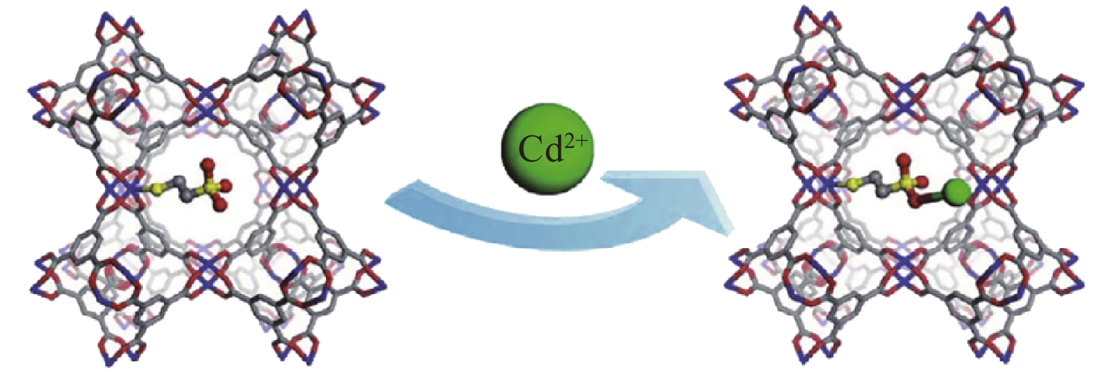
MYD1-72-Gas6 Complex Crystal Structure
4.2 Preclinical Data
In ovarian cancer PDX models, MYD1-72 monotherapy achieved:
- Primary tumor volume reduction: 95% (vs. PBS control).
- Metastasis count: Decreased from 28 to 3 (P<0.001).
- Combination with doxorubicin: 78% complete remission rate (CR), with significantly lower cardiotoxicity than conventional chemotherapy.
5. Challenges and Future Directions
5.1 Delivery System Innovation
Current MYD1 drug limitations include:
- Short plasma half-life (t1/2 = 2.3 hours) requiring frequent dosing.
- Low tumor penetration (<8% injected dose reaches lesions).
Solutions include: - PEGylation: Extends circulation time to 18 hours.
- pH-responsive nanoparticles: Enable tumor-specific drug release in acidic microenvironments.
5.2 Combination Therapy Strategies
Preliminary data show MYD1-72 synergizes with PD-1 antibodies:
- CD8+ T-cell infiltration: Increased 4.7-fold.
- Immunosuppressive Treg proportion: Reduced from 22% to 7%.
Conclusion
As a molecular bridge linking innate and adaptive immunity, MYD1's functional polymorphism offers unique therapeutic perspectives. Integrating protein engineering with nanotechnology, MYD1-72-based biologics could overcome existing treatment barriers, ushering in a new era of precision immunotherapy.
This article is reviewed and published by the technical expert team of UA
Disclaimer: This article partially utilizes artificial intelligence assistance in its creation. If any content involves copyright or intellectual property issues, please let us know and we promise to verify and remove it as soon as possible.
Purchase recombinant protein, choose Nanjing UA-Bio
UA protein focuses on providing various protein reagents, raw materials, and services required for drug research and development, cell therapy, gene therapy, and basic scientific research, including drug target proteins, immune checkpoint proteins, cytokines, tool enzymes, customized protein expression, and full-length transmembrane protein development. Youai is committed to providing customers with high-quality products and professional services, and building a High-tech Biological Enterprise with International Competitiveness.







 Business license (three certificates in one)