




TNF-α: Core Target and Signaling Pathway Analysis for Autoimmune Disease Treatment
Author: Senior Product Manager of UA Views:286 


This article focuses on the molecular characteristics and biological functions of tumor necrosis factor α (TNF-α), systematically elaborating its central role as an inflammatory cytokine in immune responses and inflammation regulation, and analyzing the mechanistic differences between the signaling pathways mediated by its two receptors, TNFR1 and TNFR2.
- Recent Advances
- Product Information
Recent Advances
TNF-α: Core Therapeutic Target and Signaling Pathway Analysis in Autoimmune Diseases
Summary
This article focuses on the molecular characteristics and biological functions of tumor necrosis factor alpha (TNF-α), systematically elaborating its central role as an inflammatory cytokine in immune response and inflammation regulation, and analyzing the mechanistic differences between the signaling pathways mediated by its two receptors, TNFR1 and TNFR2.
This article focuses on the molecular characteristics and biological functions of tumor necrosis factor alpha (TNF-α), systematically elaborating its central role as an inflammatory cytokine in immune response and inflammation regulation, and analyzing the mechanistic differences between the signaling pathways mediated by its two receptors, TNFR1 and TNFR2.
1. Molecular Characteristics and Sources of TNF-α.
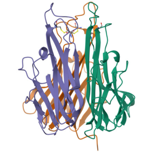
Tumor necrosis factor alpha (TNF-α) is a homotrimeric transmembrane protein composed of 157 amino acid residues, with a molecular weight of approximately 17 kDa (soluble form) to 26 kDa (transmembrane form). The human TNF-α gene is located on chromosome 6p21.3, within the major histocompatibility complex (MHC) class III gene region, and its encoded protein plays a central regulatory role in inflammatory responses. The TNF family includes TNF-α and TNF-β, which share only about 30% sequence homology but utilize the same receptor system. TNF-α accounts for 70% to 95% of total TNF biological activity and is the primary focus of research and clinical applications.
TNF-α is primarily produced by activated macrophages. Additionally, various cell types, including CD4+ T cells, natural killer cells, neutrophils, mast cells, fibroblasts, and endothelial cells, can secrete TNF-α upon inflammatory stimulation. TNF-α is synthesized as a transmembrane form (mTNF-α) on the cell surface and is released as a soluble trimer (sTNF-α) after proteolytic cleavage by TNF-α converting enzyme (TACE/ADAM17), allowing it to exert systemic effects via the bloodstream.
2. Physiological Functions and Pathological Significance of TNF-α.
Under physiological conditions, TNF-α is a critical regulator of immune defense and tissue homeostasis. During early infection, TNF-α activates vascular endothelial cells to promote leukocyte recruitment to infected sites, enhances the bactericidal activity of phagocytes, and collaborates with other cytokines to eliminate pathogens. TNF-α also participates in the development of lymphoid organs and tissue repair processes.
However, excessive production of TNF-α is closely associated with the pathological processes of various autoimmune diseases. In rheumatoid arthritis, TNF-α drives synovial fibroblast proliferation and osteoclast activation, leading to joint erosion and bone destruction. In inflammatory bowel disease, TNF-α mediates intestinal mucosal barrier disruption and epithelial cell apoptosis, exacerbating local inflammatory infiltration. In psoriasis, TNF-α promotes abnormal keratinocyte proliferation and skin inflammation. Given its central pathological role in autoimmune diseases, TNF-α has become one of the most mature and widely targeted molecules in the global autoimmune disease drug market.
3. Structure and Signaling Mechanisms of TNF Receptors.
The biological functions of TNF-α depend on its binding to two cell surface receptors: TNFR1 (TNF receptor 1, CD120a, 55-60 kDa) and TNFR2 (TNF receptor 2, CD120b, 75-80 kDa). Both receptors belong to the TNF receptor superfamily, with extracellular domains rich in cysteine repeat motifs, but their intracellular domains differ significantly, leading to distinct signaling functions.
TNFR1 is widely expressed on almost all nucleated cells, with further upregulation under inflammatory conditions. The intracellular domain of TNFR1 contains a death domain (DD), which serves as the molecular basis for its apoptosis and inflammatory signaling. Upon TNF-α binding, TNFR1 trimerizes and recruits TRADD (TNFR1-associated death domain protein) to form complex I. Within complex I, TRADD further recruits TRAF2, RIPK1, and cIAPs, activating the NF-κB and MAPK pathways to promote pro-inflammatory cytokine expression and cell survival genes. After complex I dissociates, TRADD forms complex II with FADD and caspase-8, triggering apoptotic signals. Thus, TNFR1 primarily mediates pro-inflammatory signals and apoptosis, representing the core pathway through which TNF-α drives inflammation.
TNFR2 expression is more restricted, primarily found on immune cells such as regulatory T cells, monocytes, and endothelial cells. The intracellular domain of TNFR2 lacks a death domain and instead recruits TRAF2 and TRAF1 to activate the PI3K/AKT pathway, promoting cell survival, proliferation, and tissue repair. Under chronic inflammatory conditions, TNFR2's pro-inflammatory effects can also become prominent, demonstrating its context-dependent functionality. Due to differences in affinity, soluble TNF-α preferentially binds TNFR1, while transmembrane TNF-α exhibits higher affinity for TNFR2. This distinction provides a theoretical basis for selective therapeutic strategies targeting specific receptors or TNF-α forms.
4. Conclusion.
As a core inflammatory cytokine in autoimmune diseases, TNF-α mediates a complex balance between inflammation regulation, cell survival, and apoptosis through signaling networks involving TNFR1 and TNFR2. A deeper understanding of TNF-α's structural features, receptor selectivity, and signaling pathway preferences is crucial for developing safer and more effective therapeutic strategies for autoimmune diseases. Recombinant human TNF-α protein, as a vital tool for basic research and drug development, will continue to provide essential support for advancements in this field.
In TNF-α-related basic research and drug screening, high-quality recombinant human TNF-α protein is a key tool for receptor binding analysis, cellular function studies, and signaling pathway exploration. To meet these research needs, UniLove offers TNF-α Protein, Human, suitable for TNFR1/TNFR2 binding analysis, NF-κB signaling pathway investigation, and in vitro activity evaluation of anti-TNF-α antibody drugs.
This article is reviewed and published by the technical expert team of UA
Disclaimer: This article partially utilizes artificial intelligence assistance in its creation. If any content involves copyright or intellectual property issues, please let us know and we promise to verify and remove it as soon as possible.
Purchase recombinant protein, choose 
UA protein focuses on providing various protein reagents, raw materials, and services required for drug research and development, cell therapy, gene therapy, and basic scientific research, including drug target proteins, immune checkpoint proteins, cytokines, tool enzymes, customized protein expression, and full-length transmembrane protein development. Nanjing Univ is committed to providing customers with high-quality products and professional services, and building a High-tech Biological Enterprise with International Competitiveness.
Target proteins | membrane proteins | cytokines | enzymes | viral antigens | protein customization
Nanjing UNIV Biotechnology Co., Ltd. Email:order@ua-bio.com Phone:+86-25-56221161

Product Information

Link:





©2021-2026 all rights reserved UNIV Biotechnology Co.,Ltd.
Privacy Policy
网站备案号 苏ICP备2025207586号-2 浙公网安备33010202004867号
 Business license (three certificates in one)
 Tel:+86-25-56221161
E-mail:order@ua-bio.com
All products sold on this website shall not be used for clinical diagnosis or treatment of humans or animals, and shall only be used for non-medical purposes such as industrial or scientific research.


