




H-2Kd MHC Tetramer Reveals Mechanisms of Immune Evasion Related to the "Relative Rarity" Dormancy of Disseminated Tumor Cells and the Reversal Role of T Cell Immunotherapy


Though anti - tumor immune responses are activated, dormant disseminated tumor cells (DTCs) can still survive long - term. The specific mechanisms of how they escape immune surveillance remain unclear. This article introduces a study published in Cancer Cell Metabolism (IF = 50.3) in January 2025. It reveals a newly - recognized immune evasion paradigm - the "relative rarity" effect - which is the pathological basis for the persistence of DTCs.
- Recent Advances
- Product Information
Immune evasion of dormant disseminated tumor cells is due to their scarcity and can be overcome by T cell immunotherapies
Despite activated antitumor immune responses, dormant disseminated tumor cells (DTCs) persist long-term. The mechanisms underlying their immune evasion remain poorly understood. This study, published in Cancer Cell Metabolism (IF=50.3) in January 2025, uncovers a novel immune evasion paradigm termed "relative scarcity," which forms the pathological basis of DTC persistence. Notably, T cell-based immunotherapies may inadvertently trigger adaptive exhaustion mechanisms in DTCs by increasing interaction frequency between DTCs and antigen-specific T cells. These findings provide a new perspective on nonlinear interactions within the tumor immune microenvironment and offer theoretical frameworks for optimizing existing immunotherapeutic strategies.

Highlights
- First identification of "relative scarcity" as the core mechanism of DTC immune evasion.
- A critical threshold exists for DTC-T cell interaction frequency; low-frequency contacts fail to trigger elimination.
- Enhancing T cell-DTC interaction frequency breaks immune tolerance, enabling DTC eradication.
- Vaccination or adoptive T cell therapy emerges as a potential strategy to eliminate minimal residual disease.
Research Background
Epidemiological evidence indicates that late-stage tumor relapse often originates from early dissemination of tumor cells to peripheral tissues like lungs and bone marrow. In preclinical breast cancer models, approximately 30% of patients exhibit single DTCs in bone marrow aspirates, most of which are Ki67⁻ (non-proliferative). Preclinical studies confirm that DTC clearance correlates with metastasis-free survival: reducing DTC burden improves outcomes in murine models and breast cancer patients. However, immunotherapeutic targeting of DTCs is hindered by difficulties in identifying specific markers, slowing clinical translation.
The mechanisms of DTC-host immune interaction remain incompletely understood. Some studies suggest DTCs may evade surveillance by mimicking tissue stem cell quiescence, including downregulating MHC class I (MHC I)—critical for CD8⁺ T cell recognition. Restoring MHC I expression has been proposed to enhance immune detection, but clinical data showing T cell recognition of DTCs challenges this hypothesis.
Chimeric antigen receptor (CAR) T cell therapy offers new hope for DTC targeting. Unlike MHC I-dependent natural T cells, CAR-T cells directly kill antigen-expressing cells, achieving remarkable success in hematologic malignancies. Clinical trials demonstrate CAR-T efficacy in eliminating minimal residual disease in bone marrow. However, solid tumors remain refractory due to immunosuppressive microenvironments. Whether similar limitations apply to DTCs is unclear, underscoring the need to dissect DTC immune evasion mechanisms.
Key Findings
1. Persistence of dormant DTCs despite functional antigen-specific T cells
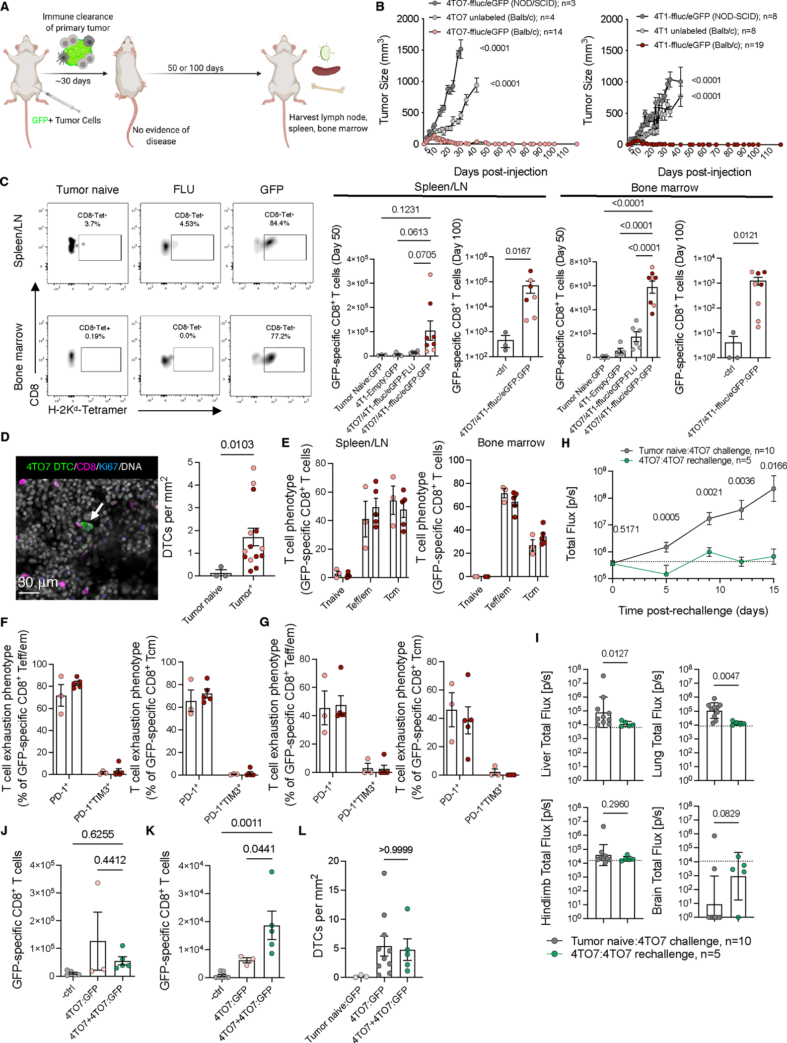
Even after complete pathologic remission of primary tumors, immune checkpoint inhibitors fail to eliminate residual DTCs. To investigate barriers to DTC clearance, researchers tested whether DTCs expressing dominant neoantigens could be recognized by endogenous tumor-specific T cells. Balb/c mice inoculated with parental/wild-type or ffluc/eGFP⁺ allogeneic breast tumor cells rejected primary tumors but retained eGFP⁺ DTCs in bone marrow (confirmed by confocal imaging, Fig 1A-D). Despite CD8⁺ T cell infiltration (tetramer staining, Fig 1C), DTCs persisted.
Phenotypic analysis revealed eGFP-specific memory T cells with effector (CD44ᴴⁱCD62Lˡᵒ) and central memory (CD44ᴴⁱCD62Lʰⁱ) profiles (Fig 1E). While these cells expressed PD-1 (comparable to tetramer⁻ cells), they lacked exhaustion markers like TIM-3 (Fig 1F-G), suggesting retained surveillance capacity. Bioluminescent imaging (BLI) confirmed resistance to tumor rechallenge (Fig 1H-I), with increased eGFP-specific T cell frequencies post-rechallenge (Fig 1J-K). However, DTC numbers remained unchanged (Fig 1L), indicating unresolved immune tolerance.
2. DTCs evade immunity via MHC I downregulation
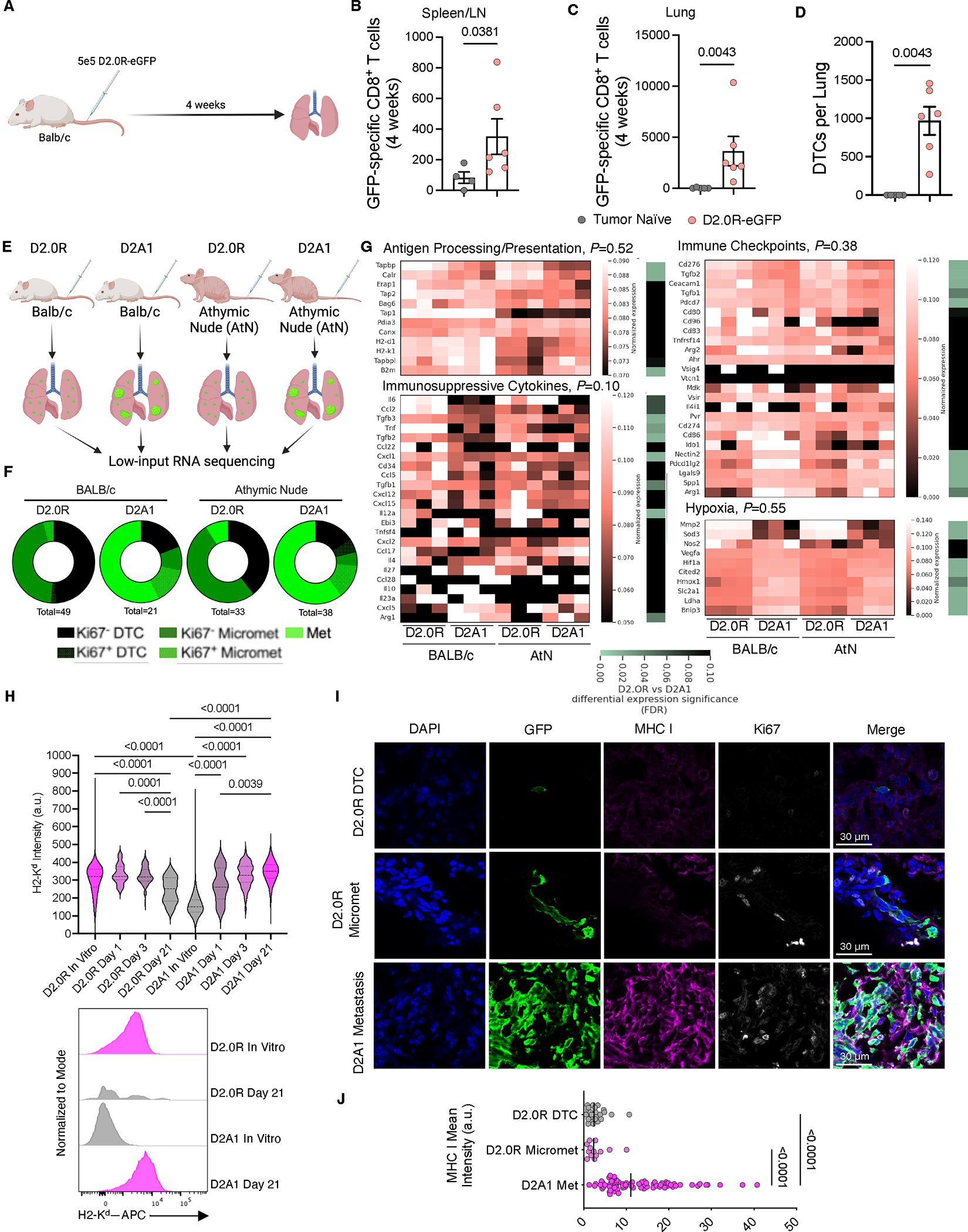
Using intravenous D2.0R-eGFP breast tumor cells in Balb/c mice, researchers observed long-term (4–240 days) pulmonary DTC persistence in quiescent (Ki-67⁻) states (Fig 2A-D). Despite thousands of antigen-specific T cells in lungs (Fig 2C), DTCs persisted. Low-input RNA sequencing of DTCs from immunocompetent and T cell-deficient (AtN) mice revealed no significant differences in antigen processing, immunosuppressive factors, or checkpoint genes, but reduced hypoxia signatures (Fig 2E-G). Notably, D2.0R cells progressively downregulated MHC I (H-2Kᵈ), unlike proliferative D2A1 cells (Fig 2H-J). This downregulation occurred independently of proliferation (Ki-67 status) or adaptive immune pressure, confirming MHC I loss as a core evasion mechanism.
3. MHC I-restricted T cells effectively eliminate dormant DTCs
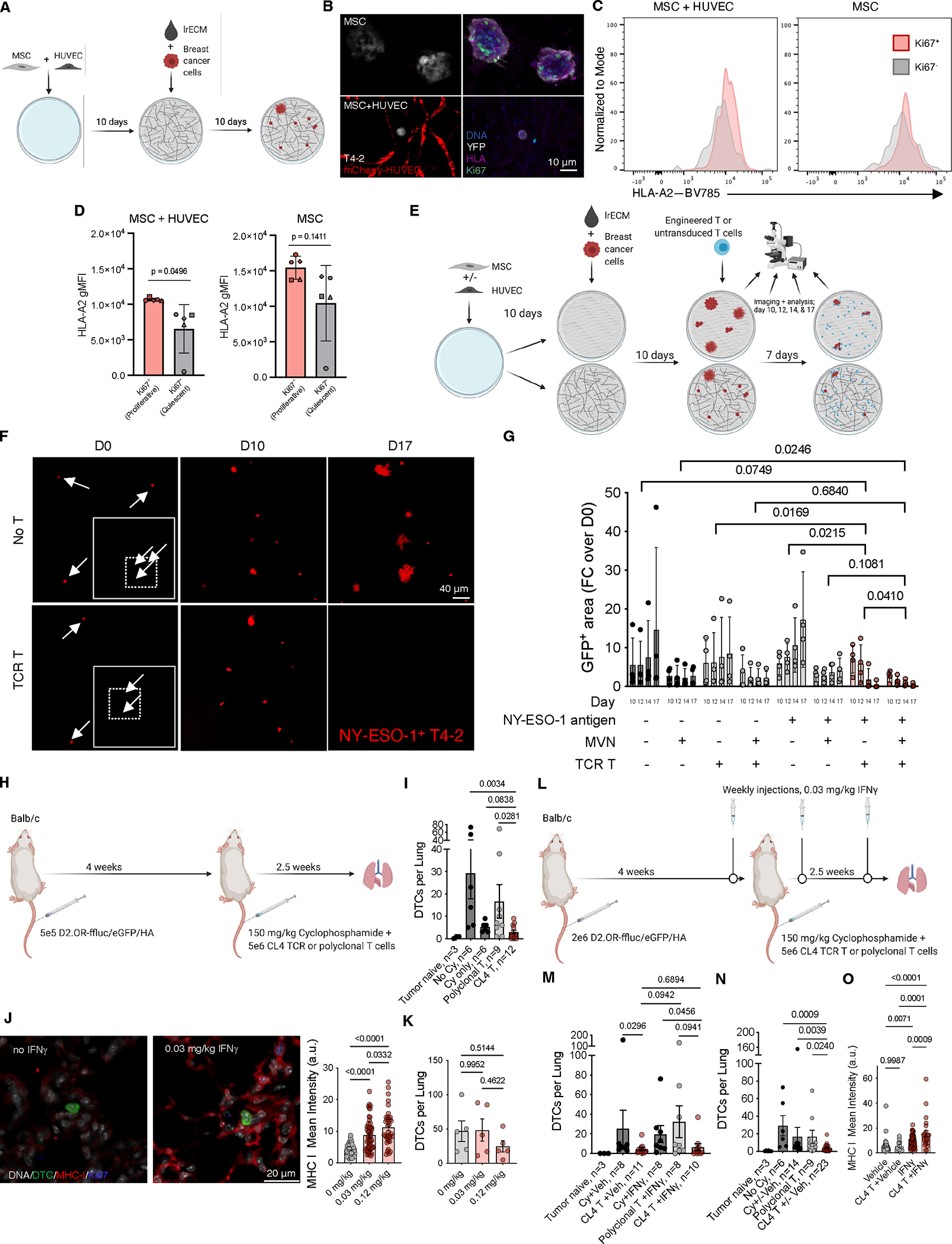
A microvascular niche (MVN) 3D culture system induced T4-2 breast cancer cell quiescence, mirroring in vivo MHC I downregulation (Fig 3A-D). HLA-A2/NY-ESO-1-specific TCR-T cells killed 84–97% of NY-ESO-1⁺ T4-2 cells, irrespective of proliferation status (Fig 3E-G). In vivo, CL4 TCR-T cells cleared pulmonary D2.0R DTCs more effectively than polyclonal T cells or chemotherapy (Fig 3I). Low-dose IFNγ upregulated DTC MHC I (Fig 3J) but did not enhance endogenous responses (Fig 3K) or TCR-T efficacy (Fig 3L-N), suggesting MHC I-independent resistance in residual DTCs (Fig 3O).
4. Relative scarcity underpins DTC immune evasion
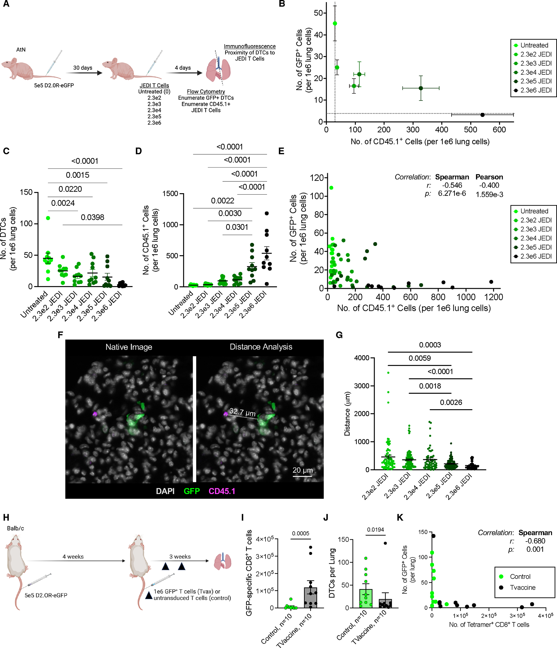
Adoptive transfer of JEDI TCR-T cells into D2.0R-bearing AtN mice revealed dose-dependent DTC clearance (Fig 4A-D). Increased T cell density reduced DTC-T cell spatial separation (Fig 4F-G), correlating with enhanced killing. Tumor vaccination expanded eGFP-specific CD8⁺ T cells 18-fold (Fig 4I), achieving 53% DTC clearance (Fig 4J). A treatment-resistant case with low T cell infiltration further emphasized the critical role of T cell quantity (Fig 4I-K).
5. CAR-T cells overcome relative scarcity for efficient DTC clearance
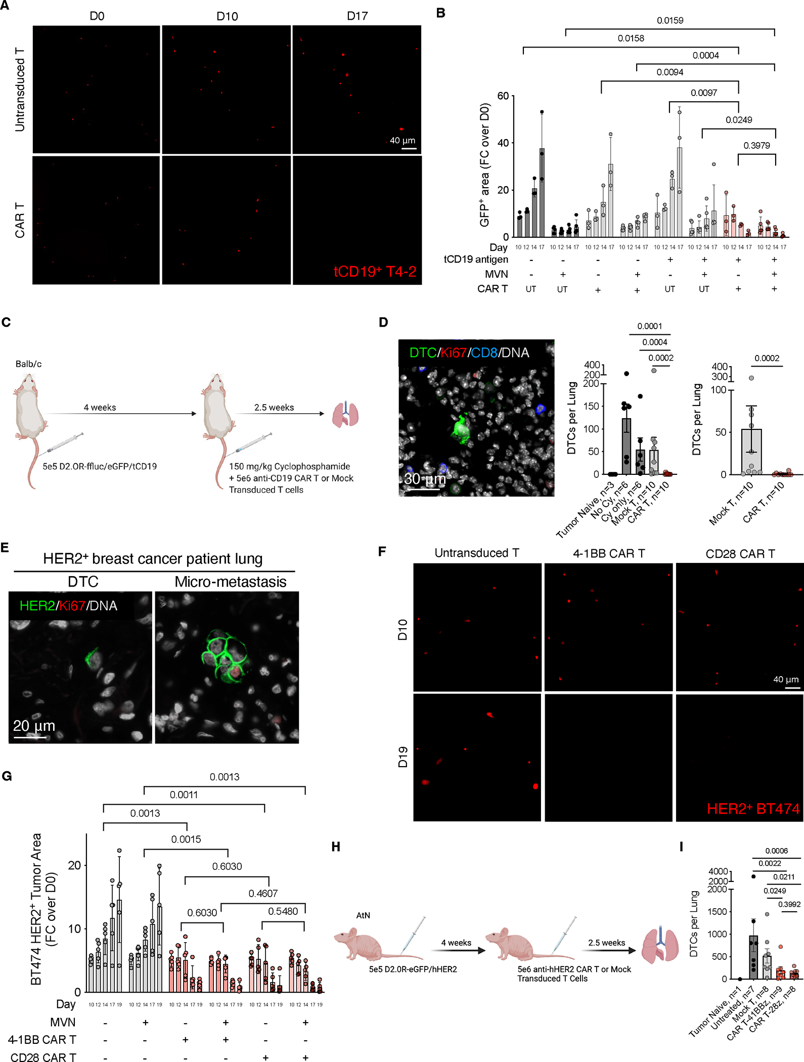
In 3D cultures, anti-tCD19 CAR-T cells eliminated 93% of proliferative/quiescent T4-2 cells (Fig 5A-B). In vivo, anti-CD19 CAR-T cells cleared 98% of pulmonary D2.0R DTCs (Fig 5C-D). HER2-specific CAR-T cells reduced HER2⁺ BT474/HCC1569 lesions by 89–97% in immunodeficient mice (Fig 5E-I), confirming MHC-independent efficacy.
Conclusion
This study identifies "relative scarcity" as a novel immune evasion mechanism limiting DTC-T cell interactions. Breaking this threshold requires increasing effector T cell density to enhance interaction frequency. Comparative analysis of TCR-T cells, CAR-T cells, and vaccines highlights the therapeutic potential of elevating effector-to-target ratios. These findings provide a rationale for targeting high-immunogenicity DTC antigens and engineering T cell therapies to overcome minimal residual disease. Future studies must explore microenvironmental reprogramming by immunotherapies to advance precision oncology.
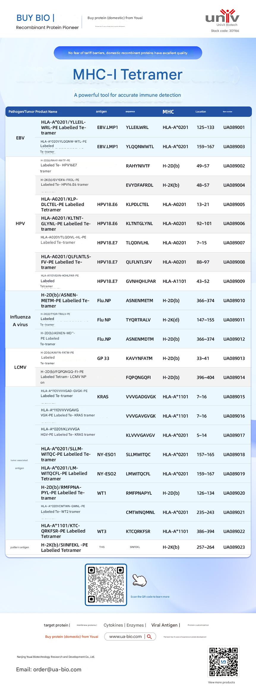
| Disease Category | Product Name | Antigen | Sequence | MHC | Position | Product Number |
|---|---|---|---|---|---|---|
| EBV | HLA-A*0201/YLELLVWRL-PE Labelled Tetramer | EBV.LMP1 | YLELLVWRL | HLA-A*0201 | 125-133 | UA089001 |
| EBV | HLA-A*0201/YLQQNWTL-PE Labelled Tetramer | EBV.LMP1 | YLQQNWTL | HLA-A*0201 | 159-167 | UA089003 |
| EBV | H-2Db(b)/RAHY-NIVTF-PE Labelled Tetramer | HPV16.E7 | RAHYNIVTF | H-2Db | 49-57 | UA089002 |
| HPV | H-2K(b)/EVYDFA-FRQL-PE Labelled Tetramer | HPV16.E6 | EVYDFARDL | H-2Kb | 48-57 | UA089004 |
| HPV | HLA-A*0201/KLP-DLCTL-PE Labelled Tetramer | HPV18.E6 | KLPDCTL | HLA-A*0201 | 13-21 | UA089005 |
| HPV | HLA-A*0201/KLTNT-GLYQL-PE Labelled Tetramer | HPV18.E6 | KLTNTGLYNL | HLA-A*0201 | 92-101 | UA089006 |
| HPV | HLA-A*0201/TLODIVIHL-PE Labelled Tetramer | HPV18.E7 | TLODIVIHL | HLA-A*0201 | 7~15 | UA089007 |
| HPV | HLA-A*0201/QFLNTL-FV-PE Labelled Tetramer | HPV18.E7 | QFLNTLFSV | HLA-A*0201 | 88-97 | UA089008 |
| HPV | HLA-A*1101/GVNHQLPAR-PE Labelled Tetramer | HPV18.E7 | GVNHQLPAR | HLA-A*1101 | 43-52 | UA089009 |
| Influenza A Virus | H-2D(b)/ASNENMETM-PE Labelled Tetramer | Flu.NP | ASNENMETM | H-2Db | 366-374 | UA089010 |
| Influenza A Virus | H-2K(d)/TYQR-TRALY-PE Labelled Tetramer | Flu.NP | TYQRTRALY | H-2Kd | 147-155 | UA089011 |
| Influenza A Virus | H-2D(b)/ASNEN-MDTM-PE Labelled Tetramer | Flu.NP | ASNENMDTM | H-2Db | 366-374 | UA089012 |
| LCMV | H-2D(b)/KAVYNFATM-PE Labelled Tetramer | GP 33 | KAVYNFATM | H-2Db | 33-41 | UA089013 |
| LCMV | H-2D(b)/FQPGQGFVK-PE Labelled Tetramer | LCMV NP | FQPGQGFVK | H-2Db | 396-404 | UA089014 |
| Tumor-related | HLA-A*1101/VVGADGVK-PE Labelled Tetramer | KRAS | VVGADGVK | HLA-A*1101 | 7~16 | UA089015 |
| Tumor-related | HLA-A*1101/VVGAGVGK-PE Labelled Tetramer | KRAS | VVGAGVGK | HLA-A*1101 | 7~16 | UA089016 |
| Tumor-related | HLA-A*0201/KLVVGAGV-PE Labelled Tetramer | KRAS | KLVVGAGV | HLA-A*0201 | 5~14 | UA089017 |
| Tumor-related | HLA-A*0201/SLLMWITQC-PE Labelled Tetramer | NY-ESO1 | SLLMWITQC | HLA-A*0201 | 157-165 | UA089018 |
| Melanoma | HLA-A*0201/LMWITQCFL-PE Labelled Tetramer | NY-ESO2 | LMWITQCFL | HLA-A*0201 | 159-167 | UA089019 |
| Melanoma | H-2Db(b)/MMFPNA-P1-PE Labelled Tetramer | WT1 | RMFPNAPL | H-2Db | 126-134 | UA089020 |
| Melanoma | HLA-A*0201/CMTWV-PE Labelled Tetramer | WT2 | CMTWVNMDM | HLA-A*0201 | 235-243 | UA089021 |
| Melanoma | HLA-A*1101/KTCQRKSF-PE Labelled Tetramer | WT3 | KTCQRKSF | HLA-A*1101 | 386-394 | UA089022 |
| Ovarian Cancer | H-2K(b)/SINFEKL-PE Labelled Tetramer | OVA | SINFEKL | H-2Kb | 257-264 | UA089023 |
This article is reviewed and published by the technical expert team of UA
Disclaimer: This article partially utilizes artificial intelligence assistance in its creation. If any content involves copyright or intellectual property issues, please let us know and we promise to verify and remove it as soon as possible.
Purchase recombinant protein, choose Nanjing UA-Bio
UA protein focuses on providing various protein reagents, raw materials, and services required for drug research and development, cell therapy, gene therapy, and basic scientific research, including drug target proteins, immune checkpoint proteins, cytokines, tool enzymes, customized protein expression, and full-length transmembrane protein development. Youai is committed to providing customers with high-quality products and professional services, and building a High-tech Biological Enterprise with International Competitiveness.







 Business license (three certificates in one)