LCMV MHC Tetramer Technology
The activation of antigen-specific (AgS) T cells offers an innovative way to overcome current T cell therapy limitations. This study developed two immunotherapeutic platforms—Immuno-STAT and Neo-STAT—that can selectively control tumor-specific immune responses. By delivering co-stimulatory ligands and peptide-HLA (pHLA) complexes precisely to AgS T cells, these platforms showed great potential. In particular, IL-2 variants with optimized affinity, when deployed on this platform, demonstrated specific expansion of oligoclonal and multifunctional AgS T cells in vitro. They also synergized with CD80 signaling, significantly outperforming traditional peptide stimulation in terms of proliferation efficiency.
Clinical Translation Bottlenecks of Adoptive T Cell Therapy (ACT)
ACT has made significant clinical progress by activating and genetically modifying patients' T cells outside the body. However, its broad application is hindered by complex cell preparation, strict reinfusion requirements, individualized patient preconditioning, and potential safety risks. Systemic co-stimulation strategies aim to directly activate tumor-specific T cells within patients. However, existing approaches (e.g., anti-CD137 antibodies combined with high-dose IL-2) have been limited by severe toxicities like vascular leak syndrome and cytokine storms.
The Efficacy-Toxicity Paradox of IL-2 Therapy
IL-2, a classic immunomodulatory factor, can induce the proliferation and differentiation of CD8+ effector T cells (Teff), NK cells, and B cells. It has achieved partial remission in metastatic renal cell carcinoma and melanoma. However, its clinical application is severely limited by dose-dependent toxicities (e.g., vascular leak syndrome) and the risk of expanding regulatory T cells (Tregs). Tregs, which highly express the high-affinity IL-2 receptor (CD25), proliferate preferentially under IL-2 stimulation, suppressing Teff's anti-tumor effects and directly correlating with poor cancer prognoses. Additionally, while CTLA-4 and PD-1 inhibitors significantly enhance T cell responses, their combined toxicity limits this application.
Targeted Delivery Strategies to Overcome Immunotherapy Dilemmas
Scalable therapies like cancer vaccines show potential but rely heavily on the integrity of antigen-presenting cells (APCs). Dendritic cells (DCs), key APCs, can initiate anti-tumor CD8+ T cell responses by presenting pHLA and co-stimulatory molecules like CD80/CD86. However, their function is significantly weakened by inhibitory receptors (e.g., PD-L1, TIM-3) and immunosuppressive factors (e.g., TGF-β, IDO) in the tumor microenvironment. Thus, developing non-APC-dependent, targeted immune modulation strategies is crucial for breaking clinical bottlenecks.
Technological Innovations of Immuno-STAT/Neo-STAT Platforms
The Immuno-STAT and Neo-STAT platforms, designed based on natural T cell regulation mechanisms, achieve precise signal delivery through an Fc fusion protein architecture (Figure 1a). Composed of peptide epitopes, MHC class I molecules, co-modulatory molecules, and Fc segments, these platforms activate homologous T cells through multivalent binding. Their modular design allows flexible fusion of co-stimulatory ligands (e.g., CD80, 4-1BBL) or co-inhibitory ligands (e.g., PD-L1) to the MHC-Fc heavy chain or β2-microglobulin (β2m) terminus, enabling precise control of the spatial conformation and stoichiometry of signaling molecules.
IL-2 Optimized Variants for Selective T Cell Expansion
Initial validation focused on IL-2 as a co-modulatory molecule. By optimizing affinity to reduce its binding to CD25 on Tregs while retaining activation of the intermediate-affinity IL-2R (CD122/CD132) on Teff, these IL-2 variants were shown to selectively expand AgS CD8+ Teff cells in vitro. They induced multifunctional effector molecules (e.g., IFN-γ, Granzyme B) and synergized with CD80 signaling, achieving a 3-5 times higher proliferation efficiency than traditional peptide stimulation.
Platform Advantages and Clinical Translation Prospects
Compared to traditional systemic immunostimulation, the Immuno-STAT/Neo-STAT platforms offer safety and efficacy enhancements through the following mechanisms:
Targeted delivery: Fc-mediated pHLA-TCR binding ensures localized enrichment of signaling molecules, reducing non-specific activation. Signal integration: Co-localization of co-stimulatory molecules and IL-2 variants forms immunological synapse-like structures, mimicking natural T cell activation. Overcoming resistance: Bypassing APC defects and tumor microenvironment inhibition, these platforms directly activate resting AgS T cells.
These platforms provide a new paradigm for developing next-generation cancer immunotherapies, with their modular design supporting rapid iterative optimization to address tumor heterogeneity and drug-resistant mutations.
Optimization and Assessment of IL-2-Based Immuno-STAT Frameworks
To optimize IL-2-based Immuno-STAT frameworks, this study systematically evaluated a series of constructs for efficacy, antigen specificity (AgS), and manufacturability. Candidate constructs, centered on the LCMV gp33-41/H-2Db epitope-MHC complex, were engineered by N-terminal fusion of human IL-2 mutants and C-terminal linkage of effector-function-attenuated mouse IgG2a Fc segments. The design strategy included two key optimizations:
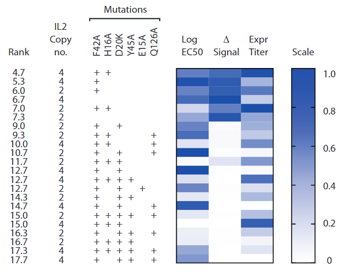
IL-2 functional modification: Introduction of IL-2 attenuating mutations (F42A/H16A) to reduce IL-2Rα-dependent toxicity and Treg binding affinity while preserving activation of the intermediate-affinity IL-2Rβ/γ receptor. Stoichiometry control: Limiting IL-2 copy numbers to 2 or 4 units to prevent protein expression titer drops from excessive fusion (Figure S2).
In Vitro Functional Validation and Screening
Humanized IL-2 variants exhibited potent activity in both human and mouse cell lines, with STAT5 phosphorylation (pSTAT5) indicating IL-2R pathway activation, strongly correlating with T cell proliferation and phenotypic marker expression. Constructs were assessed using AgS P14 TCR transgenic mice and non-AgS C57BL/6 mice splenic CD8+ T cells, evaluating:
Efficacy index: LogEC50-P14 (half-maximal effective concentration of AgS P14 T cell pSTAT5 signal). Selectivity index: ΔlogEC50 (EC50 difference between AgS P14 and non-AgS C57BL/6 cells). Manufacturability score: Based on protein expression titer and purification yield.
Preliminary screening showed LCMV-IST-IL2.FH4 performed best, followed by LCMV-IST-IL2.F4 (Figure S3). Dose-gradient validation (Figure 1b) revealed:
No significant difference in AgS efficacy: logEC50 values for inducing pSTAT5 in AgS P14 T cells were similar across constructs. Comparable non-AgS background signals: No significant difference in activating non-AgS C57BL/6 T cells (logEC50-B6). Consistent selectivity: Parameters measured by two methods showed no significant differences among constructs.
Structure-Function Relationship Analysis
Despite expectations that F42A/H16A mutations would weaken IL-2R signaling, data showed that increasing IL-2 copies (e.g., LCMV-IST-IL2.4 vs. LCMV-IST-IL2.2) did not reduce AgS efficacy or selectivity. Possible explanations include:
Multivalent binding compensation: High IL-2 copy numbers enhance local binding to AgS T cells via steric hindrance, offsetting reduced single-molecule affinity. Signal integration advantage: pMHC-TCR binding-triggered immunological synapse formation may enrich IL-2 variants on the T cell surface, amplifying bioactivity.
Overall, LCMV-IST-IL2.FH4, with balanced efficacy, selectivity, and manufacturability, was chosen as the priority candidate for subsequent studies, offering crucial design principles for next-generation tumor-targeting immunomodulators.
Functional Validation and Modular Expansion of Humanized Immuno-STAT Platform
To validate the clinical potential of the humanized Immuno-STAT platform, complexes integrating IL-2.FH4 variants, effector-function-attenuated human IgG1 Fc segments, HLA-A*0201 molecules, and model antigenic epitopes (CMV pp65₄₉₅-₅₀₃ or MART1₂₆-₃₅) were constructed (Figure 1c). By co-culturing human peripheral blood mononuclear cells (PBMCs) with specific Immuno-STAT molecules in vitro, the expansion efficiency and functional characteristics of antigen-specific CD8+ T cells were systematically assessed.
IL-2.FH4-Dependent T Cell Expansion Validation
Dual tetramer staining showed that after CMV-IST-IL2.FH4 stimulation, the frequency of CMV-specific CD8+ T cells in PBMCs significantly increased (>30-fold). In contrast, the CMV-IST group lacking the IL-2 fusion module showed no specific expansion (Figure 1d). Supplementing exogenous recombinant human IL-2 (wild-type) partially restored the expansion ability of CMV-IST, confirming that IL-2R pathway activation is essential for T cell proliferation. Similarly, MART1-IST-IL2.FH4 induced significant expansion of MART1-specific CD8+ T cells (Figure 1e), with expansion efficiency independent of antigenic epitope affinity, indicating platform compatibility with various tumor-associated antigens.
Multifunctional Effector T Cell Differentiation Characteristics
The expanded antigen-specific CD8+ T cells exhibited strong co-expression of multifunctional effector molecules:
Cytotoxic markers: >75% of cells simultaneously expressed IFN-γ, TNF-α, Granzyme B, and the degranulation marker CD107a (Figure 1f), comparable to peptide-stimulated groups. Antigen-specific activation: Under irrelevant peptide stimulation, except for Granzyme B (basal expression <5%), the expression of other effector molecules was near baseline. Dose dependence: Immuno-STAT and peptide-stimulated groups showed high consistency in functional molecule expression thresholds and dose-response curves, indicating that the induced T cells possess effect strengths similar to natural responses.
Oligoclonal TCR Lineage Fidelity
Single-cell TCR sequencing confirmed that the expanded antigen-specific CD8+ T cells maintained an oligoclonal profile consistent with peptide-stimulated groups (Figure 1g):
CMV epitope: The dominant clonotype sequences and frequency distributions were identical between the two groups. MART1 epitope: Due to low initial precursor frequencies, both showed low clonality expansion, but TCRβ chain CDR3 region sequences were highly overlapping.
Modular Platform Compatibility and Co-stimulatory Ligand Library Expansion
The Immuno-STAT platform demonstrated excellent modular adaptability:
Co-stimulatory molecule integration: In addition to IL-2, co-stimulatory ligands such as CD80, CD86, and CD137L could be flexibly fused into the MHC-Fc framework, forming a co-stimulatory signal combination library. HLA allele expansion: Complexes such as AFP₄₀₃-₄₁₁/HLA-A1101 and HBVp₁₀₉-₁₁₈/HLA-A2402 were successfully constructed, validating the platform's ability to display non-HLA-A*0201 epitopes. Low-affinity epitope processing: For tumor-associated antigens with weak HLA binding (e.g., KRAS G12D), the Neo-STAT technology was developed. Through site-specific chemical ligation, epitopes were loaded onto pre-formed "empty" HLA molecules, overcoming the limitations of genetic fusion expression (Figure 2b).
Preliminary Exploration of Co-stimulatory Signal Synergy
Using the CMV epitope as a model, the synergy between CD80 co-stimulatory molecules and IL-2.FH4 was evaluated:
Monotherapy activity: CMV-IST-CD80-2 alone failed to induce significant T cell proliferation. Combined stimulation: When CMV-IST-CD80-2 was combined with CMV-IST-IL2.FH4, T cell expansion efficiency increased 2.3-fold compared to monotherapy groups and significantly outperformed traditional peptide stimulation (Figure 2d), suggesting that co-stimulatory signal integration could further amplify therapeutic effects.
In summary, the Immuno-STAT/Neo-STAT platform, through precise spatial arrangement of signaling molecules and modular design, enables efficient and selective expansion of antigen-specific T cells. Its compatible co-stimulatory ligand library and epitope display system lay the technical foundation for developing next-generation personalized cancer immunotherapies.
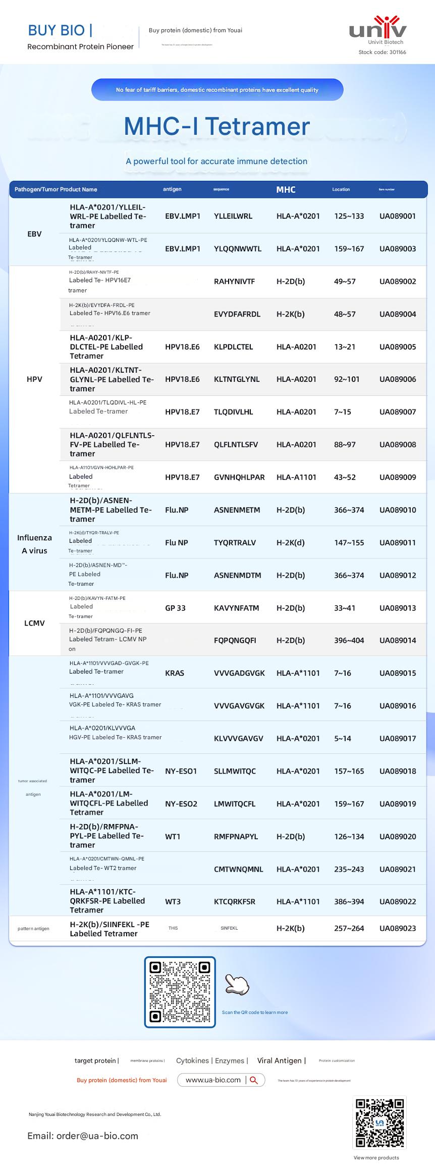
Related Products:
| Disease Category |
Product Name |
Antigen |
Sequence |
MHC |
Position |
Product Number |
| EBV |
HLA-A*0201/YLELLVWRL-PE Labelled Tetramer |
EBV.LMP1 |
YLELLVWRL |
HLA-A*0201 |
125-133 |
UA089001 |
| EBV |
HLA-A*0201/YLQQNWTL-PE Labelled Tetramer |
EBV.LMP1 |
YLQQNWTL |
HLA-A*0201 |
159-167 |
UA089003 |
| EBV |
H-2Db(b)/RAHY-NIVTF-PE Labelled Tetramer |
HPV16.E7 |
RAHYNIVTF |
H-2Db |
49-57 |
UA089002 |
| HPV |
H-2K(b)/EVYDFA-FRQL-PE Labelled Tetramer |
HPV16.E6 |
EVYDFARDL |
H-2Kb |
48-57 |
UA089004 |
| HPV |
HLA-A*0201/KLP-DLCTL-PE Labelled Tetramer |
HPV18.E6 |
KLPDCTL |
HLA-A*0201 |
13-21 |
UA089005 |
| HPV |
HLA-A*0201/KLTNT-GLYQL-PE Labelled Tetramer |
HPV18.E6 |
KLTNTGLYNL |
HLA-A*0201 |
92-101 |
UA089006 |
| HPV |
HLA-A*0201/TLODIVIHL-PE Labelled Tetramer |
HPV18.E7 |
TLODIVIHL |
HLA-A*0201 |
7~15 |
UA089007 |
| HPV |
HLA-A*0201/QFLNTL-FV-PE Labelled Tetramer |
HPV18.E7 |
QFLNTLFSV |
HLA-A*0201 |
88-97 |
UA089008 |
| HPV |
HLA-A*1101/GVNHQLPAR-PE Labelled Tetramer |
HPV18.E7 |
GVNHQLPAR |
HLA-A*1101 |
43-52 |
UA089009 |
| Influenza A Virus |
H-2D(b)/ASNENMETM-PE Labelled Tetramer |
Flu.NP |
ASNENMETM |
H-2Db |
366-374 |
UA089010 |
| Influenza A Virus |
H-2K(d)/TYQR-TRALY-PE Labelled Tetramer |
Flu.NP |
TYQRTRALY |
H-2Kd |
147-155 |
UA089011 |
| Influenza A Virus |
H-2D(b)/ASNEN-MDTM-PE Labelled Tetramer |
Flu.NP |
ASNENMDTM |
H-2Db |
366-374 |
UA089012 |
| LCMV |
H-2D(b)/KAVYNFATM-PE Labelled Tetramer |
GP 33 |
KAVYNFATM |
H-2Db |
33-41 |
UA089013 |
| LCMV |
H-2D(b)/FQPGQGFVK-PE Labelled Tetramer |
LCMV NP |
FQPGQGFVK |
H-2Db |
396-404 |
UA089014 |
| Tumor-related |
HLA-A*1101/VVGADGVK-PE Labelled Tetramer |
KRAS |
VVGADGVK |
HLA-A*1101 |
7~16 |
UA089015 |
| Tumor-related |
HLA-A*1101/VVGAGVGK-PE Labelled Tetramer |
KRAS |
VVGAGVGK |
HLA-A*1101 |
7~16 |
UA089016 |
| Tumor-related |
HLA-A*0201/KLVVGAGV-PE Labelled Tetramer |
KRAS |
KLVVGAGV |
HLA-A*0201 |
5~14 |
UA089017 |
| Tumor-related |
HLA-A*0201/SLLMWITQC-PE Labelled Tetramer |
NY-ESO1 |
SLLMWITQC |
HLA-A*0201 |
157-165 |
UA089018 |
| Melanoma |
HLA-A*0201/LMWITQCFL-PE Labelled Tetramer |
NY-ESO2 |
LMWITQCFL |
HLA-A*0201 |
159-167 |
UA089019 |
| Melanoma |
H-2Db(b)/MMFPNA-P1-PE Labelled Tetramer |
WT1 |
RMFPNAPL |
H-2Db |
126-134 |
UA089020 |
| Melanoma |
HLA-A*0201/CMTWV-PE Labelled Tetramer |
WT2 |
CMTWVNMDM |
HLA-A*0201 |
235-243 |
UA089021 |
| Melanoma |
HLA-A*1101/KTCQRKSF-PE Labelled Tetramer |
WT3 |
KTCQRKSF |
HLA-A*1101 |
386-394 |
UA089022 |
| Ovarian Cancer |
H-2K(b)/SINFEKL-PE Labelled Tetramer |
OVA |
SINFEKL |
H-2Kb |
257-264 |
UA089023 |












 Business license (three certificates in one)