




Deciphering MHC I Multimer Technology: A Revolution from Molecular Design to Immune Monitoring


Cytotoxic T lymphocytes (CTLs), serving as core effector cells in immune responses, execute critical immune surveillance functions through the specific recognition of antigens via their T cell receptors (TCRs). During antigen recognition, the CD8 co-receptor of CTLs collaborates with TCRs to precisely identify antigenic peptides presented by MHC class I molecules.
- Recent Advances
- Product Information
MHC I Multimer Technology
Do you know the types of MHC I multimers? What are the differences between Tetramers, Pentamers, and Streptamers?
In the article "Deciphering the World of MHC I Multimers" published by IBA Lifesciences, the types, differences, characteristics, and development prospects of MHC multimers are summarized. The full text can be freely downloaded at the end of this article.
MHC Multimers: Revolutionary Tools for Immunological Research
Cytotoxic T lymphocytes (CTLs), serving as core effector cells in immune responses, execute critical immune surveillance functions through the specific recognition of antigens via their T cell receptors (TCRs). During antigen recognition, the CD8 co-receptor of CTLs collaborates with TCRs to precisely identify antigenic peptides presented by MHC class I molecules.
MHC class I molecules are widely expressed on the surface of all nucleated cells, with their core function being to bind and display peptides derived from the degradation of endogenous cellular proteins, including viral proteins and tumor-associated antigens. Even if these peptides originate from self-proteins, once recognized as abnormal antigens by MHC class I molecules, CTLs with corresponding TCRs can initiate cytotoxic effects to precisely eliminate infected or cancerous cells, thereby effectively curbing infection spread and tumor progression.
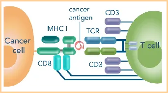
Each T cell receptor (TCR) performs immune surveillance functions by specifically recognizing unique peptide-MHC (pMHC) complexes. To cope with the vast array of antigenic variations in the environment, the immune system must maintain a vast repertoire of TCR diversity. This diversity is primarily achieved through genetic combinatorial mechanisms: during T cell development, V(D)J gene segment rearrangement, random excision or addition of nucleotides at junctional regions, and random pairing of αβ heterodimeric chains collectively establish the foundation for TCR diversity. Theoretical calculations suggest that these mechanisms can generate up to 10^20 unique TCR molecules, providing the organism with nearly unlimited antigen-recognizing potential.
A comprehensive analysis of immune response mechanisms and an in-depth understanding of their biological foundations require systematic research on T cells and their T cell receptors (TCRs) involved in immune recognition. This research field not only belongs to the realm of basic science but is also directly relevant to applied areas such as vaccine development. For example, dynamic analysis of T cell subsets following viral infection cycles or vaccination can reveal the quality, duration, and protective characteristics of immune responses, providing key indicators for evaluating the efficacy of immune interventions.
To overcome the bottlenecks in studying antigen-specific T cells, Professor Altman's research group pioneered the technical scheme of using peptide-MHC class I (pMHC I) complexes as molecular probes in 1996. The core innovation lies in assembling multiple pMHC I monomers on a fluorescently labeled scaffold structure through chemical cross-linking to form multivalent complexes. This design significantly enhances the binding affinity of the probe to TCRs, enabling successful direct staining and flow cytometric analysis of HIV-specific T cells. This study laid the theoretical foundation for MHC multimer technology, demonstrating that low-affinity pMHC I monomers can be functionally enhanced through spatial aggregation effects to stably bind TCRs on target cell surfaces. After nearly three decades of development, MHC multimer technology has become the standard tool for studying antigen-specific T cells, continuously driving the mechanistic analysis and clinical translational research of adaptive immune responses.
Types of MHC I Multimers
Tetramers
The first MHC class I multimer technology developed in 1996 is known as MHC I tetramers. Its core structure consists of four peptide-MHC class I (pMHC I) complexes covalently bound to an avidin scaffold. Avidin, a tetrameric glycoprotein derived from egg white, contains a high-affinity biotin-binding site in each subunit, capable of simultaneously binding four biotin-labeled pMHC I monomers. As an alternative, streptavidin—a non-glycosylated avidin analog derived from Streptomyces—has gradually become the preferred scaffold for tetramer construction due to its lower non-specific binding background. By covalently coupling fluorescent dyes to the scaffold protein, tetramer complexes enable direct flow cytometric staining of antigen-specific T cells. The advent of tetramer technology has laid the foundation for the development of MHC multimer tools and continues to drive the optimization of antigen-specific T cell detection techniques. In addition to classical tetramers, currently commercialized MHC class I multimer products also include Pentamers, Streptamers, and Dextramers, each exhibiting differentiated advantages in scaffold design, binding kinetics, and experimental application scenarios. Furthermore, novel multimer technologies such as NTAmers are also under continuous development, further enriching the technological spectrum of MHC multimers.
Pentamers
MHC class I Pentamer technology emerged in 2000, featuring an innovative structure composed of five pMHC I complexes covalently assembled on a self-assembling coiled-coil scaffold. This coiled-coil domain, derived from the natural polymerization module of cartilage oligomeric matrix protein (COMP), enables directional arrangement of five pMHC I molecules through genetic engineering, ensuring that all antigen-binding sites are spatially oriented in the same direction. This design significantly enhances epitope density, allowing Pentamers to exhibit approximately 10-fold higher affinity for TCRs compared to traditional tetramers. Compared to tetramer technology, Pentamers can simultaneously conjugate five fluorescent groups or biotin labels, resulting in a 40%-60% increase in fluorescence signal intensity per unit area. This enhances labeling efficiency and detection sensitivity in low-frequency antigen-specific T cell assays, particularly for rare cell populations such as tumor neoantigen- or viral variant-specific T cells.
Streptamers
MHC class I Streptamer technology, developed based on the Strep-tag® system, achieved its first commercial application in 2002. This technology employs a genetic fusion strategy to couple the C-terminus of the pMHC I molecule with the Twin-Strep-tag®, enabling it to bind with high affinity (Kd≈10⁻⁸ M) to the Streptavidin mutant Strep-Tactin®. Its core innovation lies in the introduction of a competitive dissociation mechanism: the addition of free biotin enables the high-affinity biotin to competitively displace and reversibly dissociate the pMHC I molecules, making Streptamer complexes the first reversible MHC class I multimer tool. This feature provides unique advantages in T cell sorting, in vitro functional assays, and in vivo adoptive transfer therapy research, effectively avoiding sustained TCR signaling activation caused by traditional multimer staining.
Dextramers
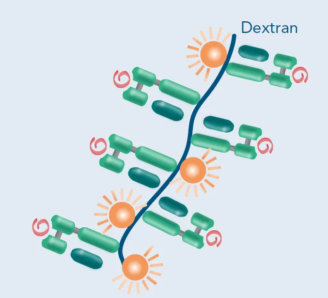
MHC class I Dextramer technology is a second-generation multimer system developed following Streptamer, named after its dextran scaffold structure. This technology forms a high-density epitope presentation platform by chemically cross-linking multiple pMHC I molecules to linear dextran fibers. The branched structure of the dextran scaffold allows each complex to load up to 50 pMHC I molecules, significantly enhancing epitope presentation efficiency. Experimental data indicate that Dextramer technology exhibits 10-100-fold higher detection sensitivity for low-affinity TCRs (dissociation rate constant Kd>10⁻⁵ M) compared to traditional tetramers, successfully enabling direct detection of rare cell populations such as tumor neoantigen-specific T cells and filling the technological gap in low-affinity epitope research.
NTAmers
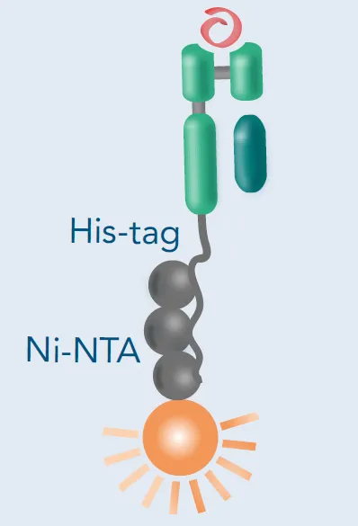
NTAmer technology represents the third generation of reversible MHC class I multimer development, named after the nickel-nitrilotriacetic acid (Ni²+-NTA) chelation system. This technology achieves multimerization by genetically introducing a 6× histidine tag (His-tag) into the pMHC I complex and utilizing Ni²+-NTA chelation with histidine. Its dissociation mechanism is similar to Streptamer technology: the addition of imidazole competitively binds to Ni², leading to the dissociation of metal chelation bonds and subsequent multimer dissociation. Although no commercial NTAmer products are currently available, preliminary studies suggest that this system holds potential applications in TCR-pMHC interaction kinetics research and long-term live-cell imaging. Its adjustable dissociation rate provides a novel tool for dissecting the spatiotemporal regulation of TCR signaling.
Reversibility: A Key Feature of MHC Multimers
Reversibility serves as a distinguishing characteristic among different MHC class I multimer technological systems. Traditional MHC multimers (such as Tetramers and Pentamers) rely on covalent cross-linking or high-affinity non-covalent interactions to achieve multimerization of pMHC I molecules, significantly enhancing epitope density and TCR binding strength. However, this irreversible binding mode may lead to negative effects such as staining complex internalization, T cell functional inhibition, or apoptosis, and limits the study of TCR-ligand dissociation kinetics.
Technological Principle Comparison
Classical Tetramers rely on the avidin-biotin system (Kd≈10⁻¹⁵ M) to form ultra-stable complexes but lack controllable dissociation capabilities. Pentamers and Dextramers, while optimizing detection sensitivity through epitope density enhancement, still maintain irreversible binding characteristics. In contrast, MHC class I Streptamer technology employs the specific binding of Twin-Strep-tag® to the Streptavidin mutant Strep-Tactin® (Kd≈10⁻⁸ M), enabling controllable dissociation of pMHC I molecules through competitive biotin elution. NTAmer technology, based on the Ni²+-NTA chelation system, provides another reversible solution using imidazole-mediated His-tag dissociation.
MHC I Streptamer Cell Staining Experimental Protocol
MHC class I Streptamer reagents provide an innovative tool for antigen-specific T cell detection and sorting. Its dual-color fluorescent labeling system (Streptamer complex + anti-Strep-Tactin® fluorescent antibody) enables precise identification of rare cell populations as low as 0.001%, with a signal-to-noise ratio 5-8 fold higher than traditional tetramers.
Core advantages of this technology include:
- Label-free sorting: Stained cells can be completely de-labeled through biotin elution, avoiding fluorescence residue interference in downstream functional experiments.
- Functional integrity preservation: Dissociated T cells maintain full proliferative capacity and effector functions, directly applicable to in vitro expansion, killing assays, or adoptive transfer studies.
- Dynamic analysis compatibility: Supports real-time monitoring of TCR-pMHC interaction kinetics, providing a technological platform for immunological synapse formation mechanism research.
Preclinical studies demonstrate that tumor-infiltrating lymphocytes (TILs) sorted using Streptamer maintain a 3-fold higher antigen-specific retention rate after in vitro expansion compared to traditional methods, laying a methodological foundation for the development of personalized cellular therapy products.
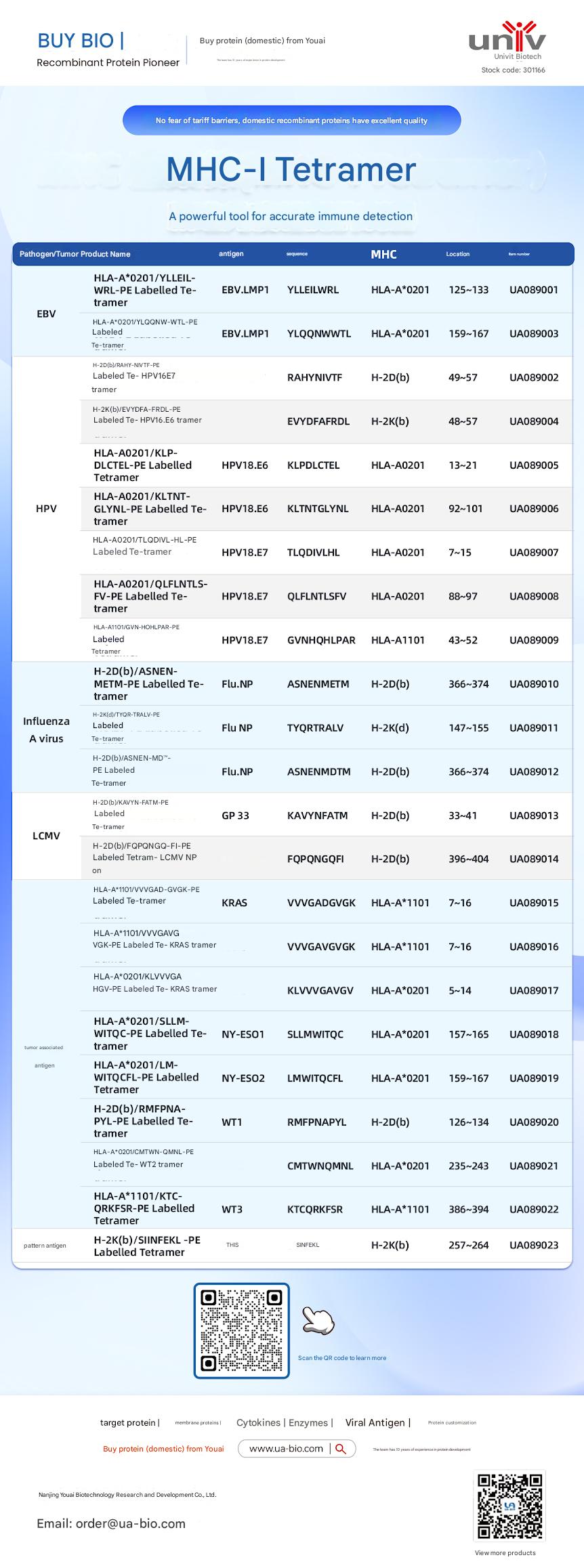
| Disease Category | Product Name | Antigen | Sequence | MHC | Position | Product Number |
|---|---|---|---|---|---|---|
| EBV | HLA-A*0201/YLELLVWRL-PE Labelled Tetramer | EBV.LMP1 | YLELLVWRL | HLA-A*0201 | 125-133 | UA089001 |
| EBV | HLA-A*0201/YLQQNWTL-PE Labelled Tetramer | EBV.LMP1 | YLQQNWTL | HLA-A*0201 | 159-167 | UA089003 |
| EBV | H-2Db(b)/RAHY-NIVTF-PE Labelled Tetramer | HPV16.E7 | RAHYNIVTF | H-2Db | 49-57 | UA089002 |
| HPV | H-2K(b)/EVYDFA-FRQL-PE Labelled Tetramer | HPV16.E6 | EVYDFARDL | H-2Kb | 48-57 | UA089004 |
| HPV | HLA-A*0201/KLP-DLCTL-PE Labelled Tetramer | HPV18.E6 | KLPDCTL | HLA-A*0201 | 13-21 | UA089005 |
| HPV | HLA-A*0201/KLTNT-GLYQL-PE Labelled Tetramer | HPV18.E6 | KLTNTGLYNL | HLA-A*0201 | 92-101 | UA089006 |
| HPV | HLA-A*0201/TLODIVIHL-PE Labelled Tetramer | HPV18.E7 | TLODIVIHL | HLA-A*0201 | 7~15 | UA089007 |
| HPV | HLA-A*0201/QFLNTL-FV-PE Labelled Tetramer | HPV18.E7 | QFLNTLFSV | HLA-A*0201 | 88-97 | UA089008 |
| HPV | HLA-A*1101/GVNHQLPAR-PE Labelled Tetramer | HPV18.E7 | GVNHQLPAR | HLA-A*1101 | 43-52 | UA089009 |
| Influenza A Virus | H-2D(b)/ASNENMETM-PE Labelled Tetramer | Flu.NP | ASNENMETM | H-2Db | 366-374 | UA089010 |
| Influenza A Virus | H-2K(d)/TYQR-TRALY-PE Labelled Tetramer | Flu.NP | TYQRTRALY | H-2Kd | 147-155 | UA089011 |
| Influenza A Virus | H-2D(b)/ASNEN-MDTM-PE Labelled Tetramer | Flu.NP | ASNENMDTM | H-2Db | 366-374 | UA089012 |
| LCMV | H-2D(b)/KAVYNFATM-PE Labelled Tetramer | GP 33 | KAVYNFATM | H-2Db | 33-41 | UA089013 |
| LCMV | H-2D(b)/FQPGQGFVK-PE Labelled Tetramer | LCMV NP | FQPGQGFVK | H-2Db | 396-404 | UA089014 |
| Tumor-related | HLA-A*1101/VVGADGVK-PE Labelled Tetramer | KRAS | VVGADGVK | HLA-A*1101 | 7~16 | UA089015 |
| Tumor-related | HLA-A*1101/VVGAGVGK-PE Labelled Tetramer | KRAS | VVGAGVGK | HLA-A*1101 | 7~16 | UA089016 |
| Tumor-related | HLA-A*0201/KLVVGAGV-PE Labelled Tetramer | KRAS | KLVVGAGV | HLA-A*0201 | 5~14 | UA089017 |
| Tumor-related | HLA-A*0201/SLLMWITQC-PE Labelled Tetramer | NY-ESO1 | SLLMWITQC | HLA-A*0201 | 157-165 | UA089018 |
| Melanoma | HLA-A*0201/LMWITQCFL-PE Labelled Tetramer | NY-ESO2 | LMWITQCFL | HLA-A*0201 | 159-167 | UA089019 |
| Melanoma | H-2Db(b)/MMFPNA-P1-PE Labelled Tetramer | WT1 | RMFPNAPL | H-2Db | 126-134 | UA089020 |
| Melanoma | HLA-A*0201/CMTWV-PE Labelled Tetramer | WT2 | CMTWVNMDM | HLA-A*0201 | 235-243 | UA089021 |
| Melanoma | HLA-A*1101/KTCQRKSF-PE Labelled Tetramer | WT3 | KTCQRKSF | HLA-A*1101 | 386-394 | UA089022 |
| Ovarian Cancer | H-2K(b)/SINFEKL-PE Labelled Tetramer | OVA | SINFEKL | H-2Kb | 257-264 | UA089023 |
This article is reviewed and published by the technical expert team of UA
Disclaimer: This article partially utilizes artificial intelligence assistance in its creation. If any content involves copyright or intellectual property issues, please let us know and we promise to verify and remove it as soon as possible.
Purchase recombinant protein, choose Nanjing UA-Bio
UA protein focuses on providing various protein reagents, raw materials, and services required for drug research and development, cell therapy, gene therapy, and basic scientific research, including drug target proteins, immune checkpoint proteins, cytokines, tool enzymes, customized protein expression, and full-length transmembrane protein development. Youai is committed to providing customers with high-quality products and professional services, and building a High-tech Biological Enterprise with International Competitiveness.







 Business license (three certificates in one)