I. Molecular Characteristics of LIF and Research Background of Cortical Development
Leukemia-inhibitory factor (LIF), an important member of the interleukin-6 cytokine family, is encoded by a gene located in a specific region of human chromosomes. It binds to the LIF receptor (LIFR) and gp130 co-receptor on the cell membrane to form a complex, activating downstream signaling pathways such as JAK-STAT, MAPK, and PI3K, which participate in various biological processes including cell proliferation, differentiation, and survival. As a multifunctional cytokine, LIF plays a key role in physiological processes such as embryonic development, immune regulation, and tissue repair, with its regulatory functions in nervous system development receiving increasing attention in recent years.
The normal development of the cerebral cortex depends on the precise regulation of neural stem cells. Radial glial (RG) cells, as the main neural precursor cells, their balance between proliferation and differentiation is crucial for cortical structure formation. Outer radial glial cells (oRG), an important subtype of RG, significantly expand in number during human cortical development and participate in cortical lamination and neural network construction by generating neurons and glial cells. Studies have found that oRG cells highly express LIFR during neurogenesis, suggesting that the LIF signaling pathway may be involved in regulating oRG cell fate determination, although its specific mechanism of action has not been fully elucidated.
II. Expression Characteristics of LIF Signaling Pathway in Neural Cells
(I) Cell-Specific Expression Pattern
Analysis of cortical tissues using immunofluorescence co-staining showed that the LIF receptor LIFR co-localizes with the radial glial cell marker NESTIN in the dorsal cortical region, indicating that LIF signaling is mainly activated in cortical radial glial cells. Further studies revealed that LIFR expression is region-specific, primarily distributed in oRG cells of the dorsal cortex, while showing low expression levels in other neural regions such as the ventral ganglionic eminence. Analysis of single-cell RNA sequencing databases confirmed that LIFR exhibits a dynamic expression pattern during cortical development, with the highest expression in oRG cells during the peak period of neurogenesis.
The expression of LIF ligand also shows spatiotemporal specificity. During the neurogenic stage of cortical development, LIF secretion levels significantly increase and reach an expression peak, coinciding with the active proliferation period of oRG cells. This spatiotemporal matching of ligand and receptor expression provides a structural basis for the LIF signaling pathway to regulate oRG cell functions, suggesting that it may play an important regulatory role during critical periods of cortical development.
(II) Cell Type-Specific Signal Activation
Functional studies indicated that cortical oRG cells are the main cell type responding to LIF signals. By constructing a cortical organoid model and detecting signaling pathway activity, it was found that after LIF treatment, the phosphorylation level of STAT3 in oRG cells significantly increased, while no obvious signal activation was observed in other neural precursor cell types. This cell type-specific signal response may be related to the high expression of LIFR and downstream signaling molecules in oRG cells, enabling them to specifically receive and transmit LIF signals, thereby regulating their own biological behaviors.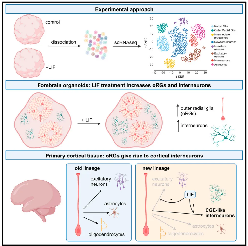
III. Functional Regulation of LIF Signaling Pathway in Cortical Development
(I) Impact on oRG Cell Proliferation and Self-Renewal
Studies using cortical organoid models confirmed that activation of the LIF signaling pathway significantly regulates the proliferation ability of oRG cells. After adding recombinant LIF protein to primary neural culture cortical organoids, the number of oRG cells significantly increased, and the expression levels of their specific markers Pax6 and HOPX were significantly upregulated. Cell cycle analysis showed that LIF treatment shortened the G1 phase and increased the proportion of S phase cells in oRG cells, indicating that LIF signaling can promote the proliferation cycle of oRG cells and maintain their self-renewal ability.
Conversely, treatment of organoids with LIF signaling pathway inhibitors led to a significant reduction in oRG cell number, accompanied by increased expression of cell differentiation markers, suggesting that inhibiting LIF signaling promotes oRG cells to exit the proliferative state and initiate differentiation programs. These results indicate that the LIF signaling pathway is a key regulatory factor maintaining oRG cell proliferation capacity, participating in cortical development by balancing proliferation and differentiation.
(II) Regulatory Role in Neuronal Generation
In addition to affecting oRG cell proliferation, the LIF signaling pathway also participates in cortical neuron generation by regulating cell fate determination. Studies found that activating the LIF signaling pathway can significantly increase the number of interneurons. In cortical organoids, LIF treatment significantly increased the expression levels of interneuron markers GABA, DLX2, and SST, while no significant changes were observed in excitatory neuron markers, indicating that LIF signaling has a selective regulatory effect on neuron subtype generation.
Lineage tracing experiments further confirmed that after LIF signal activation, the differentiation ratio of oRG cells into interneurons significantly increased, suggesting that LIF may promote interneuron generation by inducing fate bias in oRG cells. This finding reveals the important role of the LIF signaling pathway in regulating cortical neuron subtype diversity, providing a new perspective for understanding the formation mechanism of complex neural networks in the human cortex.
IV. Mechanism of Action and Research Significance of LIF Signaling Pathway
(I) Molecular Regulatory Mechanism
Mechanistic studies showed that the LIF signaling pathway exerts regulatory effects by activating the STAT3 transcription factor. In oRG cells, after LIF binds to the LIFR/gp130 complex, it recruits JAK kinase to phosphorylate STAT3, enabling it to enter the nucleus and bind to the promoter regions of downstream target genes. ChIP-sequencing analysis found that STAT3 can directly regulate the expression of cell cycle-related genes (such as Cyclin D1) and neurodifferentiation-related genes (such as NeuroD1), maintaining oRG cell characteristics and regulating their differentiation into interneurons by promoting the expression of proliferation-related genes and inhibiting the transcription of differentiation-related genes.
In addition, LIF signaling can also crosstalk with other signaling pathways (such as the Notch pathway) to synergistically regulate oRG cell fate determination. This synergistic effect of signaling networks may be an important mechanism ensuring precise regulation of cortical development.
(II) Research Value and Prospects
Research on the regulation of cortical development by the LIF signaling pathway provides important insights into understanding the formation mechanism of the human cerebral cortex. Given the key role of oRG cells in human cortical expansion, clarifying the regulatory mechanism of LIF signaling on oRG cells helps reveal the molecular basis of human cortical evolution. In terms of clinical translation, this research provides a new direction for studying neurodevelopmental disorders, as abnormalities in the LIF signaling pathway may be related to the occurrence of cortical developmental disorders, and targeted regulation of LIF signaling may become a potential therapeutic strategy for related diseases.
Future research can further explore the interaction between the LIF signaling pathway and other cortical development regulatory factors, clarify its functional differences in cortical development across different species, and simulate human cortical development processes through more precise organoid models to deeply 解析 the spatiotemporal regulatory network of LIF signaling, providing a more solid theoretical basis for neurodevelopmental biology research and related disease treatment.












 Business license (three certificates in one)