1. Molecular Characteristics of Siglec-7 and Basis of Ligand Recognition
As a member of the sialic acid-binding immunoglobulin-like lectin (Siglecs) family, which are immune receptors dependent on glycan recognition, Siglec-7 has attracted much attention due to its potential value in cancer immune regulation. Siglec-7 belongs to the CD33-related rapidly evolving subfamily and is a type I transmembrane protein. Its extracellular region contains one V-set immunoglobulin domain responsible for sialic acid binding and two C2-set immunoglobulin domains, while the intracellular region contains typical immunoreceptor tyrosine-based inhibitory motifs (ITIM) and immunoreceptor tyrosine-based switch motifs (ITSM). When bound to sialic acid-containing ligands, these motifs are phosphorylated, recruiting phosphatases such as SHP1 and SHP2, thereby inhibiting downstream immune activation signals. This mechanism is similar to that of other inhibitory Siglec family members.
Although the immune regulatory role of Siglec-7 has been initially confirmed, the specific characteristics of its physiological ligands have long been incompletely clarified. Recent studies through cell-based genomic screening and glycomic analysis have revealed the ligand recognition pattern of Siglec-7: this receptor has high specificity for the O-linked tetrasaccharide structure "disialyl core 1", and this binding depends on the protein scaffold where the ligand is located. Among them, the N-terminal region of the cell surface mucin-type glycoprotein CD43, which is rich in adjacent clusters of disialyl core 1 O-glycans, becomes a key binding target of Siglec-7. In addition, mucin-type glycoproteins such as PSGL1 and GP1α have also been confirmed to serve as ligands for Siglec-7, but their affinity and binding efficiency are lower than those of CD43, indicating that the protein scaffold structure of the ligand has an important impact on the recognition efficiency of Siglec-7.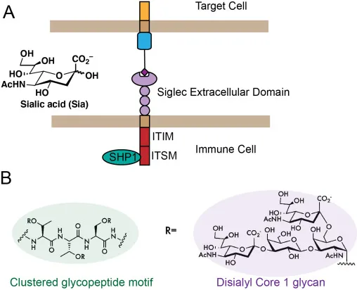
Figure 1: A) Siglec immune receptor binds to polysaccharide ligands. Then, signals are transduced through the intracellular ITIM/ITSM domain. B) Chemical structure of Siglec-7 binding motif
2. Expression Characteristics of Siglec-7 Ligands in Peripheral Immune Cells
To clarify the cellular distribution pattern of Siglec-7 ligands, researchers conducted a systematic analysis of different immune subsets in human peripheral blood mononuclear cells (PBMCs). Through staining with recombinant Siglec-7-Fc protein combined with flow cytometry detection, it was found that Siglec-7 ligands are highly and uniformly expressed on the surface of CD3⁺ T cells, while only weak and partially positive staining signals are observed in CD3⁻ cells (such as CD14⁺ monocytes and CD19⁺ B cells). This result suggests that peripheral T cells are the main expressing cells of Siglec-7 ligands.
Further subset analysis showed that the expression of Siglec-7 ligands has T cell subset specificity: in CD4⁺ T cells, the ligand expression level in naïve (CD45RO⁻) cells is significantly higher than that in memory (CD45RO⁺) cells; in the functional subsets of CD4⁺ memory T cells, the ligand expression in TH1 (CXCR3⁺) cells is higher than that in TH2 (CCR4⁺CCR6⁻) and TH17 (CCR4⁺CCR6⁺) cells. It is worth noting that the activation state of T cells can dynamically regulate the expression of Siglec-7 ligands. After stimulation with CD3/CD28 antibodies and IL-2 for 5 days, the level of Siglec-7 ligands on the surface of T cells significantly decreases, while the ligand expression of the related receptor Siglec-9 significantly increases. This indicates that the biosynthesis of Siglec ligands is selectively regulated by the cell activation state, rather than the overall change in sialic acid expression.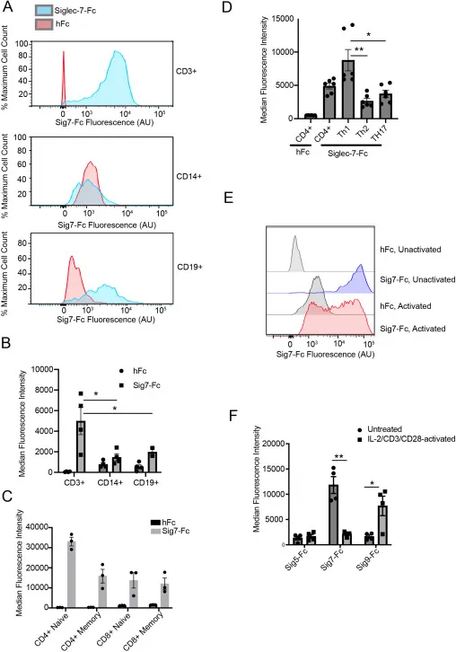
图2:具有代表性的流式细胞术结果以及不同供体的平均中值荧光强度(MFI)
3. Molecular Nature and Regulatory Mechanism of Siglec-7 Ligands
To clarify the molecular type of Siglec-7 ligands, researchers adopted a strategy combining glycan-degrading enzyme treatment and proteomic analysis, confirming that Siglec-7 ligands on peripheral immune cells are mainly O-linked glycoproteins rather than N-linked glycoproteins. Specifically, after treating PBMCs with the O-glycoproteinase StcE, the binding ability of Siglec-7-Fc significantly decreases, and its effect is comparable to that of sialidase treatment; while treatment with the N-glycoproteinase PNGase F has no significant effect on binding, clarifying the core role of O-linked glycans in Siglec-7 recognition.
Through Siglec-7-Fc protein pull-down combined with mass spectrometry analysis, CD43 was identified as the ligand with the strongest binding ability to Siglec-7 in PBMCs, and its enrichment degree is more than 50 times that of other candidate proteins. Further studies found that CD43 expressed by T cells presents a specific glycoform of 125 kDa, which is rich in disialyl core 1 O-glycans; while CD43 expressed by monocytes is mainly a 150 kDa glycoform, and its glycan structure is mostly sialylated core 2, which cannot effectively bind to Siglec-7. Glycomic analysis confirmed that the disialyl core 1 structure is the most abundant O-glycan type on the T cell surface, accounting for more than 60% of the total O-glycan structures, which provides a structural basis for the specific interaction between Siglec-7 and T cells.
The cell-specific expression of Siglec-7 ligands is mainly regulated by the glycosyltransferase GCNT1. GCNT1 can catalyze the conversion of core 1 O-glycans to core 2 structures, and core 2 structures cannot be recognized by Siglec-7. In naïve T cells, the mRNA expression level of GCNT1 is significantly lower than that in monocytes and B cells, leading to the accumulation of core 1 O-glycans, thereby highly expressing Siglec-7 ligands; conversely, monocytes have high expression of GCNT1, resulting in an increase in the proportion of core 2 O-glycans and a decrease in Siglec-7 ligand expression. Experiments confirmed that knocking down GCNT1 in the monocytic cell line THP-1 can significantly increase the expression of Siglec-7 ligands and change the CD43 glycoform from 150 kDa to 125 kDa, further verifying the regulatory role of GCNT1 as a molecular switch in the synthesis of Siglec-7 ligands.
4. Regulatory Effect and Mechanism of Siglec-7 on T Cell Functions
Given that antigen-presenting cells (such as dendritic cells and macrophages) highly express Siglec-7, while T cells highly express its ligands, researchers 推测 that Siglec-7 may regulate T cell activation through intercellular interactions. The mixed leukocyte reaction (MLR) model showed that after treating the co-culture system with Siglec-7 blocking antibodies, the interaction between DCs and T cells is enhanced, leading to a significant increase in the secretion of cytokines such as IL-6 and IFN-γ, suggesting that the binding of Siglec-7 to its ligands can inhibit T cell activation signals.
A more innovative finding is that Siglec-7 can directly regulate T cell functions. In vitro experiments showed that co-stimulation of primary T cells with soluble Siglec-7-Fc protein and anti-CD3/CD28 antibodies can significantly inhibit T cell proliferation (with a 40% decrease in CFSE dilution rate) and change the cytokine secretion profile - IL-2 secretion decreases by 35%, while the TH2-type cytokine IL-4 secretion increases by 2.5 times. This indicates that Siglec-7 not only inhibits T cell activation but also may promote their polarization to the TH2 phenotype. This phenomenon is different from the traditional cognition that Siglec-7 only functions as an inhibitory receptor, suggesting that it may play a role by directly activating signaling pathways in T cells. This mechanism is similar to the mode by which galectins trigger T cell signals through glycoprotein aggregation, providing a new perspective for understanding the versatility of the Siglec family.
5. Research Significance and Prospects
This study systematically clarified the molecular characteristics, expression regulatory mechanism of Siglec-7 ligands, and their regulatory effect on T cell functions, and first confirmed the existence of a new Siglec-7-mediated signaling axis. This axis inhibits excessive T cell activation and participates in their phenotypic polarization under physiological conditions through the interaction of "antigen-presenting cell Siglec-7-T cell ligands". This discovery breaks through the traditional cognition that the Siglec family only regulates innate immunity through inhibitory signals and reveals its new function in adaptive immune regulation.
In terms of application value, these findings provide a theoretical basis for immune therapy targeting Siglec-7. In cancer treatment, blocking Siglec-7 may relieve its inhibition on T cells and enhance anti-tumor immune responses; while in autoimmune diseases, activating Siglec-7 signals may inhibit overactive T cells and alleviate inflammatory reactions. Future studies need to further analyze the specific signaling pathways triggered by Siglec-7 in T cells, clarify its cross-regulatory mechanism with TCR signals, and explore its therapeutic potential in different disease models to promote the development and application of Siglec-7-targeted therapies.













 Business license (three certificates in one)

