




The mechanism of action of Siglec-9 in tumor immune escape


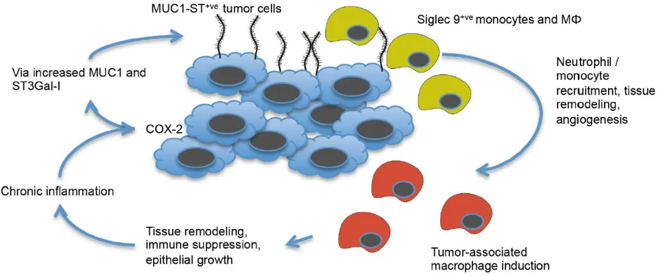
The Siglec family (sialic acid binding immunoglobulin like lectins) act as glycoprotein receptors on the surface of immune cells, participating in immune regulation by recognizing sialylated glycosides. Siglec-9 is mainly expressed on the surface of immune cells such as monocytes and macrophages, and its role in tumor immunity has not been clearly defined before.
- Recent Advances
- Product Information
I. Research Background and Scientific Questions
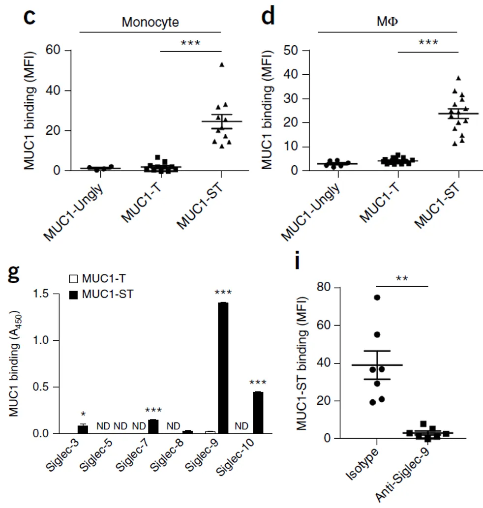
II. Specific Binding Between Abnormally Sialylated MUC1 and Siglec-9
MUC1-ungly: containing only the protein backbone without glycosylation modification;
MUC1-T: with basic glycosylation but no sialylation;
MUC1-ST: with both glycosylation and sialylation (abnormal modification).
III. Siglec-9-Mediated Functional Changes in Monocytes
The secretion of the pro-inflammatory factor IL-6 increases, participating in the formation of the inflammatory microenvironment;
The release of macrophage colony-stimulating factor (MCSF) increases, promoting the differentiation of monocytes into macrophages;
The angiogenesis-related factor PAI-1 is upregulated, providing conditions for tumor invasion and metastasis.
IV. Siglec-9-Dependent Abnormal Differentiation of Macrophages
The expression of surface co-stimulatory molecule CD86 and IL-12 was significantly downregulated;
Significantly inhibited the proliferation of CD8⁺ T cells and the expression of the activation molecule CD69;
Also inhibited the differentiation and maturation of dendritic cells (DCs).
V. Induction of Pro-Tumor Phenotype in Macrophages
The secretion of M-CSF, PAI-1, and epidermal growth factor (EGF) increases, participating in the regulation of macrophage self-renewal, tumor invasion, and cell proliferation;
Highly expresses M2-type macrophage markers CD206 and CD163;
The immune checkpoint molecules PD-L1 and IDO are upregulated, enhancing immunosuppression.
VI. Signaling Pathway Mechanism
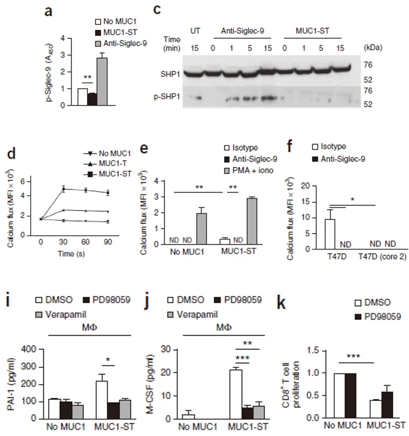
Significantly enhanced calcium ion transport in macrophages, and this effect depended on Siglec-9;
The calcium channel inhibitor verapamil could inhibit the secretion of pro-tumor factors;
Activated the MEK-ERK pathway, and MEK inhibitors could reduce the secretion of pro-tumor factors and partially restore the proliferation ability of CD8⁺ T cells.
VII. Research Significance
This article is reviewed and published by the technical expert team of UA
Disclaimer: This article partially utilizes artificial intelligence assistance in its creation. If any content involves copyright or intellectual property issues, please let us know and we promise to verify and remove it as soon as possible.
Purchase recombinant protein, choose 
UA protein focuses on providing various protein reagents, raw materials, and services required for drug research and development, cell therapy, gene therapy, and basic scientific research, including drug target proteins, immune checkpoint proteins, cytokines, tool enzymes, customized protein expression, and full-length transmembrane protein development. Nanjing Univ is committed to providing customers with high-quality products and professional services, and building a High-tech Biological Enterprise with International Competitiveness.







 Business license (three certificates in one)

