Acute-on-chronic liver failure (ACLF) is a severe liver disease syndrome characterized by systemic inflammatory response and multiple organ failure, with an extremely high mortality rate. Its core pathological features include massive hepatocyte necrosis, impaired liver regeneration capacity, and immune-metabolic disorders. How to accurately assess patient prognosis and explore effective therapeutic strategies to promote liver regeneration has always been a key issue to be solved in clinical practice.
The Syndecan family, as the main heparan sulfate proteoglycans (HSPGs) on the surface of endothelial cells and parenchymal cells, plays an important role in intercellular communication, signal transduction, and tissue repair. Among them, Syndecan-1 (SDC-1) is mainly expressed on the surface of hepatocytes and epithelial cells. Through its three heparan sulfate (HS) chains attached to the extracellular segment, it acts as a binding partner of integrins or a co-receptor for growth factors, participating in the regulation of various biological processes such as cell adhesion, migration, proliferation, and intracellular signal transduction. In the state of infection or tissue damage, SDC-1 can be shed from the cell surface into the bloodstream, and its circulating level has been confirmed to reflect the degree of vascular endothelial damage and disease severity, making it a potential prognostic marker for various severe diseases. However, the specific mechanism of action, clinical prognostic value, and potential as a therapeutic target of SDC-1 in ACLF remain unclear, and related studies provide a new research perspective for analyzing the pathophysiological process of ACLF.
Clinical cohort studies detected serum SDC-1 levels in patients with different liver diseases and found that the baseline SDC-1 concentration in ACLF patients was significantly higher than that in patients with liver cirrhosis, chronic hepatitis B, and hepatocellular carcinoma, suggesting that the abnormal increase of SDC-1 may be closely related to the acute exacerbation process of ACLF. Further correlation analysis showed that serum SDC-1 levels were positively correlated with inflammatory and liver function indicators such as total bilirubin (TBIL), aspartate transaminase (AST), and neutrophil count, and significantly correlated with classic prognostic scoring systems such as MELD score and COSSH-ACLF II score, indicating that it can comprehensively reflect the degree of liver damage and systemic inflammatory state in ACLF patients.
Analysis based on the 28-day and 90-day follow-up outcomes of patients showed that ACLF patients who died or received liver transplantation within 90 days had significantly higher baseline serum SDC-1 levels than surviving patients; serum SDC-1 levels in patients with persistent disease deterioration were also significantly higher than those in patients with improved conditions. This result suggests that serum SDC-1 levels can be used as a potential biomarker to evaluate the short-term prognosis of ACLF patients, and its dynamic changes may reflect the trend of disease progression.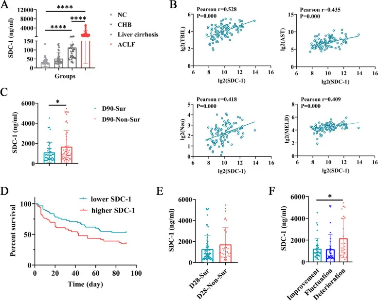
SDC-1 levels in ACLF patients and their relationship with prognosis in ACLF patients
To improve the accuracy of ACLF prognosis prediction, the study used orthogonal partial least squares discriminant analysis (OPLS-DA) to screen out the 5 most significant indicators affecting prognosis, including SDC-1, TBIL, international normalized ratio (INR), albumin, and total protein. Combined with Cox regression analysis, age, upper gastrointestinal bleeding (UGIB), SDC-1, and INR were identified as independent prognostic factors for ACLF patients, and then a new prognostic model UIAS was constructed (calculation formula: UIAS = 0.076 × age + 0.001 × SDC-1 + 1.454 × INR + 2.854 × UGIB - 12.165, where UGIB = 1 for patients without UGIB and UGIB = 2 for patients with UGIB).
Validation results showed that the UIAS score was significantly correlated with MELD, COSSH-ACLF II, and Child-Pugh scores, and showed higher efficacy in predicting the 28-day survival rate of ACLF patients (higher AUROC). Especially in patients with hepatitis B-related ACLF, the predictive AUROCs of UIAS for 28-day disease deterioration and 90-day mortality were 0.897 and 0.859, respectively, which were significantly better than the traditional COSSH-ACLF II score, confirming its practical value in clinical prognosis assessment.
To explore the pathological mechanism of SDC-1 in ACLF, the study used carbon tetrachloride (CCl4) combined with D-galactosamine (D-GalN) and lipopolysaccharide (LPS) to induce and construct a mouse ACLF model. This model showed typical ACLF characteristics: significantly increased serum ALT and AST levels, extensive hepatocyte necrosis, inflammatory cell infiltration, and liver fibrosis in liver tissue (confirmed by Masson staining), transmission electron microscopy showed hepatocyte mitochondrial swelling and disappearance of cristae structure, Tunel staining showed increased hepatocyte apoptosis, accompanied by increased infiltration of liver macrophages and neutrophils.
In this model, serum SDC-1 levels in ACLF mice were significantly increased, consistent with the performance of clinical patients; while the expression of SDC-1 in liver tissue was significantly down-regulated and distributed disorderly (SDC-1 in normal mice is mainly expressed in hepatocytes and hepatic sinusoidal endothelium), suggesting that the shedding of SDC-1 on the cell surface may be the main reason for the increase of circulating SDC-1, and its expression deficiency may be involved in the pathological process of liver injury.
Based on the biological function of SDC-1, the study selected two interventional drugs: sulodexide (SDX, which can inhibit the degradation and shedding of SDC-1) and heparan sulfate (HS, which can supplement extracellular heparan sulfate chains and promote glycocalyx repair), to evaluate their therapeutic effects on ACLF mice. The results showed that:
Improvement of liver injury: SDX and HS treatment significantly reduced hepatocyte necrosis and inflammatory infiltration in ACLF mice, mitigated mitochondrial structural damage, and decreased serum ALT and AST levels;
Inhibition of apoptosis: Tunel staining and protein detection showed that both drugs reduced hepatocyte apoptosis, down-regulated the expression of Cleaved-caspase3, and up-regulated the ratio of anti-apoptotic protein Bcl2/Bax;
Promotion of liver regeneration: SDX and HS can increase the expression of cyclin D1, cyclin E1, and proliferating cell nuclear antigen (PCNA), suggesting enhanced hepatocyte proliferation capacity.
At the same time, drug treatment can partially reverse the down-regulation of SDC-1 in liver tissue of ACLF mice and reduce serum SDC-1 levels, indicating that stabilizing the expression of SDC-1 on the cell surface or supplementing its functional fragments may be the key mechanism for improving liver injury.
To analyze the mechanism of action of SDX and HS, proteomic analysis showed that differentially expressed proteins in the SDX treatment group were enriched in cAMP, TGF-β, and MAPK signaling pathways, while differentially expressed proteins in the HS treatment group were mainly enriched in TGF-β and JAK/STAT signaling pathways. Given the known role of the JAK/STAT pathway in hepatocyte anti-apoptosis and pro-proliferation, further verification by Western blotting found that both SDX and HS could significantly increase the phosphorylation levels of JAK1 and STAT3 in the liver of ACLF mice, suggesting that they may exert liver regeneration-promoting and anti-apoptotic effects by activating the JAK1/STAT3 signaling pathway. This finding reveals the potential molecular mechanism by which SDC-1-related interventions improve ACLF, that is, by stabilizing SDC-1 expression or supplementing HS chains, activating the JAK1/STAT3 pathway, restoring hepatocyte proliferation capacity, and inhibiting apoptosis.
Existing studies have systematically clarified the dual role of SDC-1 in ACLF: as a circulating biomarker, its baseline level can effectively predict the short-term prognosis of patients, and the UIAS model constructed based on it significantly improves the accuracy of ACLF prognosis assessment; as a functional molecule, the deficiency of SDC-1 expression on hepatocyte surface is closely related to impaired liver regeneration and increased apoptosis, and interventions targeting SDC-1 (such as SDX inhibiting its shedding and HS supplementing heparan sulfate chains) can promote liver regeneration and reduce liver injury by activating the JAK1/STAT3 pathway.
These findings not only provide new biological markers and tools for the prognosis assessment of ACLF but also lay a theoretical foundation for the development of therapeutic strategies targeting SDC-1. Future studies need to further explore the specific molecular mechanism by which SDC-1 regulates the JAK1/STAT3 pathway and verify its prognostic value in larger clinical cohorts to provide more sufficient evidence for the precise diagnosis and treatment of ACLF.













 Business license (three certificates in one)

