




Cell line engineering technology based on luciferase: principles and applications in biomedical research


Luciferase can catalyze the oxidation reaction of substrate luciferin in the presence of oxygen and ATP, producing a detectable light signal without the need for external excitation light.
- Recent Advances
- Product Information
Q1: What is the fundamental principle behind using luciferase-labeled tumor cell lines in vivo?
In vivo imaging technology enables non-invasive, real-time visualization of biological processes such as tumor growth, metastasis, and gene expression in live animal models. Among various imaging modalities (e.g., ultrasound, CT, MRI, PET), bioluminescence imaging (BLI) using luciferase (Luc)-tagged cells offers distinct advantages. Luciferase catalyzes the oxidation of its substrate, luciferin, in the presence of oxygen and ATP, emitting detectable light without the need for external excitation. This reaction occurs exclusively in living cells, and light intensity correlates linearly with cell numbers. Due to minimal absorption of red light (>600 nm) by hemoglobin, emitted photons can penetrate tissues and be captured by highly sensitive cooled-CCD cameras, allowing precise quantification of tumor burden in vivo.

Q2: Does luciferase expression alter native cellular functions?
Extensive studies confirm that luciferase expression does not interfere with normal physiological activities, including cell proliferation, migration, or signaling pathways. Stable genomic integration ensures consistent expression across generations, making it a reliable tool for long-term studies.
Q3: How stable are luciferase-labeled cell lines, and is antibiotic selection necessary?
When luciferase genes are stably integrated into the host genome, expression remains consistent through cell division and differentiation. While short-term cultures may not require antibiotic selection, prolonged passaging can lead to transgene silencing or loss. Periodic antibiotic pressure or re-establishment from原始 stock is recommended to maintain signal intensity.
Q4: Why might in vivo signals appear weak or undetectable?
Several factors affect signal detection:
Instrument Sensitivity: High-performance cooled-CCD cameras and light-tight chambers are essential to capture low-intensity photons.
Substrate Kinetics: Intraperitoneal (IP) injection of luciferin yields peak emission at ~10–20 minutes, lasting ~30 minutes; intravenous (IV) injection provides faster but shorter signals. The standard dose is 150 mg/kg.
Cell Depth and Number: Signal attenuation is ~10-fold per cm depth. While subcutaneous implants may require only hundreds of cells, deep-tissue lesions need higher cell numbers.
Substrate Quality: Luciferin batches should be validated via standard curves to ensure saturation kinetics.
Q5: What are common pitfalls in luciferase assays?
Timing: Luciferase activity halves every ~30 minutes post-substrate addition; assays should be completed promptly.
Plate Selection: White opaque plates minimize cross-talk; black plates absorb signal and reduce sensitivity.
Substrate Handling: Luciferin must be stored at –20°C, protected from light, and used at saturating concentrations.
Q6: Why is hair removal critical for murine imaging?
Fur blocks, absorbs, and scatters light, while also producing autofluorescence. Depilation minimizes background noise and enhances signal-to-noise ratios.
Q7: Can deep-tissue signals be reliably detected?
Although signal intensity decreases exponentially with depth, luciferase-based detection remains effective for subcutaneous and many orthotopic models. In mice, signals can penetrate 3–4 cm, sufficient for most preclinical applications.
Q8: Beyond oncology, what are other research applications of luciferase technology?
Luciferase assays are versatile:
Stem Cell Tracking: Monitoring migration and engraftment in regenerative studies.
Infection Models: Tagging pathogens (e.g., bacteria) to study host-pathogen interactions.
Gene Regulation: Reporting promoter activity or protein-protein interactions in real time.
Drug Screening: Evaluating compound efficacy via dynamic readouts of cellular responses.
Q9: What characterizes a high-quality luciferase-labeled cell line?
Ideal lines exhibit:
Stable Genomic Integration: Ensuring consistent expression across passages.
Validated Functionality: Confirmation via STR profiling, species verification, and luciferase activity assays.
Mycoplasma-Free Status: Essential for reproducible in vivo work.
High Signal Intensity: Enabling sensitive detection in both in vitro and in vivo settings.
Example Validation:
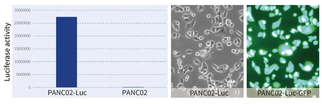
For instance, a luciferase/GFP-dual-labeled pancreatic ductal adenocarcinoma line (e.g., PANC02-Luc/GFP) allows parallel monitoring via bioluminescence (quantitative) and fluorescence (spatial), facilitating multifaceted experimental designs.
This article is reviewed and published by the technical expert team of UA
Disclaimer: This article partially utilizes artificial intelligence assistance in its creation. If any content involves copyright or intellectual property issues, please let us know and we promise to verify and remove it as soon as possible.
Purchase recombinant protein, choose Nanjing UA-Bio
UA protein focuses on providing various protein reagents, raw materials, and services required for drug research and development, cell therapy, gene therapy, and basic scientific research, including drug target proteins, immune checkpoint proteins, cytokines, tool enzymes, customized protein expression, and full-length transmembrane protein development. Youai is committed to providing customers with high-quality products and professional services, and building a High-tech Biological Enterprise with International Competitiveness.







 Business license (three certificates in one)