




Spotlight on Popular Cytokines, Part 2: GM-CSF


The colony-stimulating factor (CSF) superfamily is associated with mammalian myelopoiesis, including the development of monocytes, macrophages, dendritic cells (DCs), and polymorphonuclear macrophages. This family comprises three major members: M-CSF (macrophage colony-stimulating factor), G-CSF (granulocyte colony-stimulating factor), and GM-CSF (granulocyte-macrophage colony-stimulating factor).
- Recent Advances
- Product Information
- Reference
GM-CSF and GM-CSFR
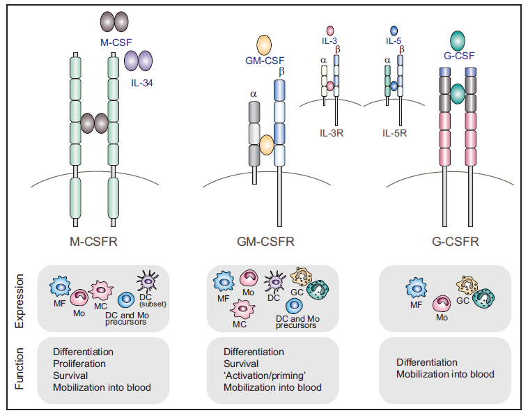
Figure 1: CSF Proteins, Receptors, and Downstream Signaling Pathways
The colony-stimulating factor (CSF) superfamily is associated with mammalian myelopoiesis, including the development of monocytes, macrophages, dendritic cells (DCs), and polymorphonuclear macrophages. This family comprises three major members: M-CSF (macrophage colony-stimulating factor), G-CSF (granulocyte colony-stimulating factor), and GM-CSF (granulocyte-macrophage colony-stimulating factor).
GM-CSF is a hematopoietic growth factor that stimulates the proliferation of myeloid cells derived from bone marrow progenitors. Human and murine GM-CSF share high homology but exhibit species-specific receptor binding. GM-CSF is named for its ability to promote the formation of eosinophil and macrophage colonies.
GM-CSF is produced by activated T cells, B cells, macrophages, endothelial cells, fibroblasts, and certain tumor cells. It plays roles in hematopoiesis, inflammatory responses, infection, monocyte and granulocyte chemotaxis, and the enhancement of cytotoxic activity.
The GM-CSF receptor (GM-CSFR) is a heterodimer composed of an α chain (CD116) and a β chain (βc), where the β chain is the signaling subunit shared with the receptors for IL-3 and IL-5. GM-CSFR is expressed on DCs, monocytes, macrophages, granulocytes, eosinophils, and endothelial cells.
Upon binding to its receptor, GM-CSF activates four major signaling pathways:
1. JAK-STAT Pathway: JAK-2 is recruited to the intracellular domain of the β chain, leading to its activation and subsequent phosphorylation of STAT5. STAT5 dimers translocate to the nucleus, promoting the expression of genes such as pim-1 and cis, which induce cell differentiation.
2. PI3K and JAK/STAT-Bcl-2 Pathways: These pathways promote cell proliferation and survival.
3. ERK1/2 and NF-κB Pathways: These pathways facilitate cell differentiation and inflammatory responses.
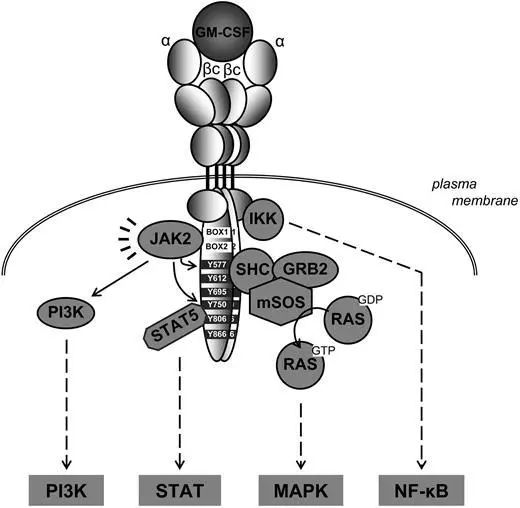
Figure 2: GM-CSF Signaling Pathway
Role of GM-CSF in Homeostasis
Epithelial cells in healthy lungs express GM-CSF. Mice deficient in GM-CSF or GM-CSFR exhibit pulmonary alveolar proteinosis (PAP), a condition characterized by the accumulation of surfactant material in alveolar spaces. PAP is rare in humans and is often associated with autoantibodies against GM-CSF or mutations in the GM-CSFR gene.
Experimental evidence suggests that PAP results from a deficiency in alveolar macrophages, which are crucial for lung homeostasis and surfactant clearance. Alveolar macrophages originate from fetal monocytes, which appear in the fetal liver by embryonic day 12.5 and subsequently colonize most embryonic tissues, including the lungs. In developing fetuses, monocytes further differentiate into immature alveolar macrophages, which mature postnatally in the alveolar spaces.
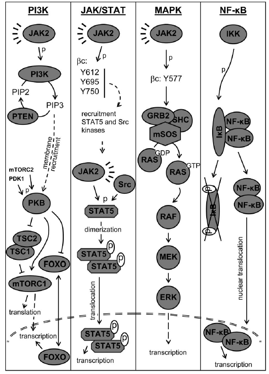
Figure 3: Role of GM-CSF in Alveolar Macrophage Function
GM-CSF was the first cytokine discovered to promote the differentiation of human monocytes or hematopoietic progenitors into DCs in vitro. These in vitro-derived DCs have been widely used in basic and clinical research.
Recent studies have revealed that bone marrow-derived DCs in mice are heterogeneous, comprising conventional DCs and monocyte-derived macrophages, both of which express MHC class II molecules and CD11c but differ significantly from in vivo DCs.
DCs are classified into three types: migratory DCs, lymphoid-resident DCs, and plasmacytoid DCs. In csf-/- or csf2rb-/- mice, migratory DCs in the skin and intestines, as well as DCs at the margins of lymphoid organs, are significantly reduced, indicating that GM-CSF is essential for DC development and maintenance.
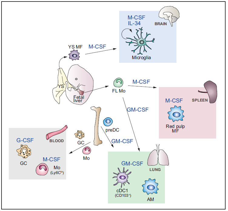
Figure 4: Effects of GM-CSF on Different Cell Types
Role of GM-CSF in Inflammation
Under inflammatory conditions, GM-CSF at the site of inflammation induces the rapid development of DC precursors from monocytes into migratory and immunologically active DCs, thereby enhancing T cell immune responses.
Th17 cells are often considered key drivers of tissue inflammation and autoimmune diseases. Studies show that the hallmark cytokine of Th17 cells, IL-17, inhibits the development of experimental autoimmune encephalomyelitis (EAE), collagen-induced arthritis (CIA), and interstitial pneumonia.

Figure 5: Role of GM-CSF in Inflammatory Responses
In models of autoimmune myocarditis, GM-CSF induces pathogenic Th17 cells by modulating the expression of IL-6 and IL-23, promoting the onset of autoimmune myocarditis. In gm-csf-/- mice, Th17 cell numbers are significantly reduced, and their activation and differentiation are impaired. Th17 and Th1 cells do not induce encephalitis, and their infiltration into the central nervous system does not cause autoimmune encephalitis. Administration of anti-GM-CSF antibodies in model mice results in mild disease, but severe autoimmune encephalitis recurs upon cessation of antibody treatment.
Additionally, researchers have found that IL-23 and IL-1β induce GM-CSF production in T cells, promoting the differentiation of Th17 and Th1/Th17 cells. Differentiated T cells produce GM-CSF, polarize macrophages toward the M1 phenotype, and induce APCs to secrete IL-6, IL-12, IL-23, and IL-1β, further enhancing GM-CSF expression.
In CIA models, gm-csf-/- mice do not develop the disease, and anti-GM-CSF antibodies are more effective than TNF antibodies in treating rheumatoid arthritis. Antibody blockade experiments demonstrate that GM-CSF is a critical mediator in pneumonia models, regulating neutrophil and macrophage proliferation and TLR4 expression.
GM-CSF not only influences macrophage and DC development but also promotes inflammatory responses and is associated with diseases such as multiple sclerosis (MS) and rheumatoid arthritis (RA).
Currently, recombinant human GM-CSF drugs are available for treating myelotoxicity induced by radiotherapy and chemotherapy, reducing the duration of neutropenia during cancer chemotherapy, and improving patient tolerance to chemotherapy.
Applications of GM-CSF:
1.Target Validation: Research in cancer and disease mechanisms.
2.Hematopoietic Stem Cell Culture: Studies in immunology.
3.ELISA Assays: Development of detection kits.
UA BIOSCIENCE offer a wide range of GM-CSF recombinant proteins from multiple species (murine and human) and expression systems. All products are activity-verified, with activities reaching 0.02 ng/mL. Some products are further validated by SPR, ELISA, and cell-based assays, ensuring comprehensive quality assurance!
Click the product code below to visit our official website for more details.
UA BIOSCIENCE GM-CSF product demonstrates comparable biological activity to competitors' products but at a more competitive price!
This article is reviewed and published by the technical expert team of UA
Disclaimer: This article partially utilizes artificial intelligence assistance in its creation. If any content involves copyright or intellectual property issues, please let us know and we promise to verify and remove it as soon as possible.
Purchase recombinant protein, choose Nanjing UA-Bio
UA protein focuses on providing various protein reagents, raw materials, and services required for drug research and development, cell therapy, gene therapy, and basic scientific research, including drug target proteins, immune checkpoint proteins, cytokines, tool enzymes, customized protein expression, and full-length transmembrane protein development. Youai is committed to providing customers with high-quality products and professional services, and building a High-tech Biological Enterprise with International Competitiveness.

1.Cytokine Signaling in Multiple Sclerosis and Its Therapeutic Applications.
2.GM-CSF as a therapeutic target in inflammatory diseases.
3.GM-CSF: From Growth Factor to Central Mediator of Tissue Inflammation.
4.Regulation of dendritic cell development by GM-CSF: molecular control and implications for immune homeostasis and therapy.






 Business license (three certificates in one)