




Unlocking T-Cell Activation: The Ultimate Switch from Dormancy to Action


As the core component of adaptive immunity, the transition of T cells from a quiescent state to a functionally activated state requires precise regulation by specific signals. This activation process constitutes a fundamental aspect of cellular immunology research and the development of immunotherapies.
- Recent Advances
- Product Information
T Cell Activation Protocol
T cells, as the core component of adaptive immunity, require precise regulation by specific signals to transition from a resting state to a functionally active state. This activation process is fundamental for cellular immunology research and the development of immunotherapies.
Part 1: The Biological Essence of T Cell Activation
T cell activation is a process of signal transduction between T cells and antigen-presenting cells via the immunological synapse structure. This process involves the reception and integration of multiple signals, ultimately leading the T cell from a quiescent state into an activated state characterized by proliferation, differentiation, and the acquisition of effector functions.
Technical Value of In Vitro Activation
-
Mechanism Studies: To dissect the regulatory roles of drugs, genes, or signaling pathways on T cell fate decisions.
-
Therapeutic Applications: To provide high-quality functional T cells for adoptive cell therapies such as CAR-T and TCR-T.
-
Immune Assessment: To evaluate vaccine efficacy or T cell function in pathological states by reconstructing immune responses in vitro.
Part 2: Core Elements – Key Signals for T Cell Activation
Complete activation of T cells requires the synergistic action of three key signals.
First Signal – Antigen-Specific Recognition
-
Molecular Mechanism: T cell receptor recognizes the MHC-peptide complex on the surface of antigen-presenting cells.
-
Technical Implementation: Anti-CD3 antibodies can specifically activate the TCR signaling pathway, mimicking the antigen recognition process.
Second Signal – Co-stimulatory Signal
-
Biological Function: Ensures the specificity of the immune response, prevents T cell anergy or apoptosis, and promotes cell proliferation and survival.
-
Technical Implementation: Anti-CD28 antibodies used in combination with anti-CD3 antibodies mimic the B7-CD28 interaction under physiological conditions.
Third Signal – Differentiation-Regulating Signal
-
Biological Function: The cytokine microenvironment determines the direction of T cell differentiation into different functional subsets.
-
Technical Implementation: IL-2, as a key growth factor, supports the clonal expansion of T cells in most culture systems.
These three signals act in a cascading and amplifying manner to collectively accomplish the complete activation and functional differentiation of T cells, providing fundamental technical support for immunological research and clinical applications.
Part 3: Mouse and Human T Cell Activation/Polarization Protocols
A. Human T Cell Activation Protocol
Protocol 1: Anti-Human CD3 Monoclonal Antibody Immobilization Method
-
Dilute the NA/LE Mouse Anti-Human CD3 monoclonal antibody in PBS to a final concentration of 5 µg/mL.
-
Add the diluted NA/LE Mouse Anti-Human CD3 monoclonal antibody to a 24-well plate at 1 mL per well for immobilization.
-
Incubate for 2 hours at 37°C with 5% CO₂, or overnight at 4°C.
-
Remove the NA/LE Mouse Anti-Human CD3 monoclonal antibody solution from the 24-well plate.
-
Prepare a single-cell suspension of human peripheral blood mononuclear cells (PBMCs). For T cell activation, cells should be resuspended in complete medium at a concentration of (1-3) × 10⁶ cells/mL. For a 24-well plate, a concentration of 1 × 10⁶ cells/mL is recommended.
-
In a V-bottom tube, prepare fully supplemented T cell medium and add the following cytokines and antibodies to the cell suspension from step 5:
-
5 µg/mL NA/LE Mouse Anti-Human CD28 monoclonal antibody
-
10 ng/mL Human Interleukin-2 (IL-2) protein
-
-
Transfer the required volume of cells to an appropriate cell culture plate, achieving a final density of (1-1.5) × 10⁶ cells/mL/cm².
-
Incubate for 2 days at 37°C with 5% CO₂.
-
After 2 days of culture, check cell health and record images.
-
Centrifuge the cell suspension at 400 × g for 5 minutes and discard the supernatant. Wash the cell pellet with PBS.
-
Centrifuge the cell suspension at 400 × g for 5 minutes and discard the supernatant. Resuspend the cells in 0.5 mL of 1% Bovine Serum Albumin (BSA) and count the cells. Assess cell viability, which should be >95%.
-
Block 1 × 10⁶ cells with 10 µg of human IgG antibody. Incubate on ice for 30 minutes.
-
Add Brilliant Violet 421™ anti-human CD25 antibody (302630) and Alexa Fluor® 488 anti-human CD69 antibody (310916) according to the manufacturer's instructions. Incubate at 4°C for 30 minutes.
-
Centrifuge the cell suspension at 400 × g for 5 minutes and wash the cell pellet twice with PBS containing 1% BSA.
-
Resuspend the cell pellet in 200 µL of PBS for flow cytometry analysis (require >10,000 cells).
Protocol 2: Anti-Human CD3 Monoclonal Antibody Soluble Method
-
Prepare a single-cell suspension of human peripheral blood mononuclear cells (PBMCs). For T cell activation, cells should be resuspended in complete medium at a concentration of (1-3) × 10⁶ cells/mL. For a 24-well plate, a concentration of 1 × 10⁶ cells/mL is recommended.
-
In a V-bottom tube, prepare fully supplemented T cell medium and add the following cytokines and antibodies to the cell suspension from step 1:
-
5 µg/mL NA/LE Mouse Anti-Human CD3 monoclonal antibody
-
5 µg/mL NA/LE Mouse Anti-Human CD28 monoclonal antibody
-
10 ng/mL Human Interleukin-2 (IL-2) protein
-
-
Transfer the required volume of cells to an appropriate cell culture plate, achieving a final density of (1-1.5) × 10⁶ cells/mL/cm².
-
Incubate for 2 days at 37°C with 5% CO₂.
-
After 2 days of culture, check cell health and record images.
-
Centrifuge the cell suspension at 400 × g for 5 minutes and discard the supernatant. Wash the cell pellet with PBS.
-
Centrifuge the cell suspension at 400 × g for 5 minutes and discard the supernatant. Resuspend the cells in 0.5 mL of 1% Bovine Serum Albumin (BSA) and count the cells. Assess cell viability, which should be >95%.
-
Block 1 × 10⁶ cells with 10 µg of human IgG antibody. Incubate on ice for 30 minutes.
-
Add Brilliant Violet 421™ anti-human CD25 antibody (302630) and Alexa Fluor® 488 anti-human CD69 antibody (310916) according to the manufacturer's instructions. Incubate at 4°C for 30 minutes.
-
Centrifuge the cell suspension at 400 × g for 5 minutes and wash the cell pellet twice with PBS containing 1% BSA.
-
Resuspend the cell pellet in 200 µL of PBS for flow cytometry analysis (require >10,000 cells).
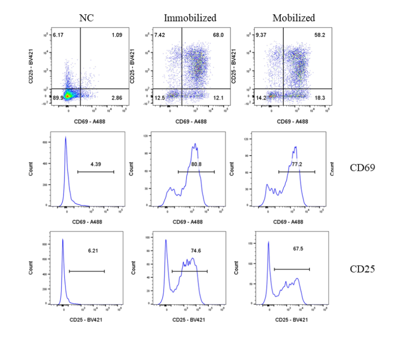 人T细胞激活试剂盒/CellXViva Human T cell Activation Kit_UA090033_优爱(UA BIOSCIENCE)官网
B. Mouse T Cell Activation Protocol
Protocol 1: Anti-Mouse CD3 Monoclonal Antibody Immobilization Method
Mouse T Cell Growth Medium Nutrient System:
-
RPMI 1640 Medium, ATCC Modification (with L-glutamine, sodium pyruvate, and HEPES)
-
12% Premium Fetal Bovine Serum
-
1% Antibiotic-Antimycotic
-
Cell Freezing: Immunocyte Serum-Free Freezing Medium
Note: The above medium system is for reference only.
-
Dilute the NA/LE Rat Anti-Mouse CD3 Recombinant mAb in PBS to a final concentration of 5 µg/mL.
-
Add the diluted NA/LE Rat Anti-Mouse CD3 Recombinant mAb to a 24-well plate at 1 mL per well for immobilization.
-
Incubate for 2 hours at 37°C with 5% CO₂, or overnight at 4°C.
-
Remove the NA/LE Rat Anti-Mouse CD3 Recombinant mAb solution from the 24-well plate.
-
Prepare a single-cell suspension from mouse spleen. For T cell activation, cells should be resuspended in complete medium at a concentration of (1-3) × 10⁶ cells/mL. For a 24-well plate, a concentration of 1 × 10⁶ cells/mL is recommended.
-
In a V-bottom tube, prepare fully supplemented T cell medium and add the following cytokines and antibodies to the cell suspension from step 5:
-
5 µg/mL NA/LE Syrian Hamster Anti-Mouse CD28 mAb
-
10 ng/mL IL-2 Protein, Mouse
-
-
Transfer the required volume of cells to an appropriate cell culture plate, achieving a final density of (1-1.5) × 10⁶ cells/mL/cm².
-
Incubate for 2 days at 37°C with 5% CO₂.
-
After 2 days of culture, check cell health and record images.
-
Centrifuge the cell suspension at 400 × g for 5 minutes and discard the supernatant. Wash the cell pellet with PBS.
-
Centrifuge the cell suspension at 400 × g for 5 minutes and discard the supernatant. Resuspend the cells in 0.5 mL of 1% Bovine Serum Albumin (BSA) and count the cells. Assess cell viability, which should be >95%.
-
Block 1 × 10⁶ cells with 10 µg of mouse IgG antibody. Incubate on ice for 30 minutes.
-
Add FITC Rat Anti-Mouse CD25 Antibody (S-R441) (S0B5215) according to the manufacturer's instructions. Incubate at 4°C for 30 minutes.
-
Centrifuge the cell suspension at 400 × g for 5 minutes and wash the cell pellet twice with PBS containing 1% BSA.
-
Resuspend the cell pellet in 200 µL of PBS for flow cytometry analysis (require >10,000 cells).
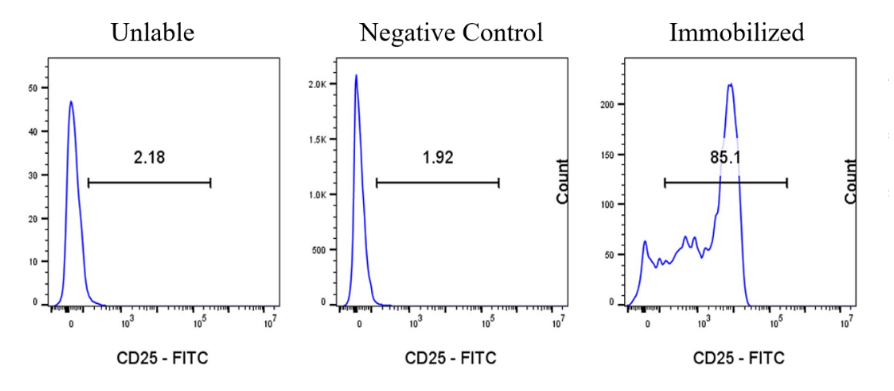
小鼠T细胞激活试剂盒/CellXViva Mouse T cell Activation Kit_UA090032_优爱(UA BIOSCIENCE)官网
Mouse T Cell Activation Kit / CellXViva Mouse T cell Activation Kit_UA090032_ UA BIOSCIENCE Official Website
This article is reviewed and published by the technical expert team of UA
Disclaimer: This article partially utilizes artificial intelligence assistance in its creation. If any content involves copyright or intellectual property issues, please let us know and we promise to verify and remove it as soon as possible.
Purchase recombinant protein, choose Nanjing UA-Bio
UA protein focuses on providing various protein reagents, raw materials, and services required for drug research and development, cell therapy, gene therapy, and basic scientific research, including drug target proteins, immune checkpoint proteins, cytokines, tool enzymes, customized protein expression, and full-length transmembrane protein development. Youai is committed to providing customers with high-quality products and professional services, and building a High-tech Biological Enterprise with International Competitiveness.







 Business license (three certificates in one)