




The Third Highlight of Best-Selling Cytokines: IL-21!


Interleukin-21 (IL-21) is primarily produced by activated CD4+ T cells, with other IL-21-expressing cells including NKT cells, Th17 cells, and CD8+ T cells.
- Recent Advances
- Product Information
- Reference
IL-21 and Its Receptor
Interleukin-21 (IL-21) is primarily produced by activated CD4+ T cells, with other IL-21-expressing cells including NKT cells, Th17 cells, and CD8+ T cells.
IL-21 consists of 162 amino acids, including four 14 kDa helical bundles. Human IL-21 shares 63% sequence identity with murine IL-21. IL-21 is a multifunctional type I cytokine that can act on various cell types, including but not limited to CD4+ T cells, CD8+ T cells, B cells, macrophages, monocytes, and dendritic cells (DCs).
The IL-21 receptor is a heterodimer composed of IL-21R (CD360) and the common cytokine receptor γ-chain (CD132, shared by IL-2, IL-4, IL-7, IL-9, and IL-15).
CD360 is a 75 kDa transmembrane glycoprotein with an intracellular domain containing six tyrosine residues, which are involved in the IL-21 signaling pathway. CD132 is a 347-amino acid protein with a molecular weight of 40 kDa.
Mutations in the human γ-chain can lead to X-linked severe combined immunodeficiency (X-SCID), a condition characterized by the absence of T cells and natural killer (NK) cells, as well as the presence of normal numbers of non-functional B cells.
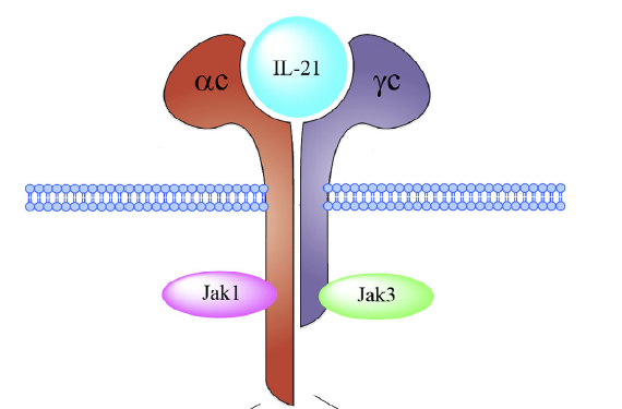
Figure 1: IL-21 and Its Receptor
Upon binding to its receptor, IL-21 induces the phosphorylation of JAK1 and JAK3. The activated JAKs then phosphorylate STAT3, STAT1, STAT5a, and STAT5b. Phosphorylated STAT3 forms dimers that translocate to the nucleus and bind to IFN-γ-activated GAS sequences, regulating IL-21-related gene expression, including Bcl-2, BAD (anti-apoptotic gene), AICDA (activation-induced cytidine deaminase), IFNG, SOCS3 and SOCS1 (suppressors of cytokine signaling 1 and 3), and IL-21.
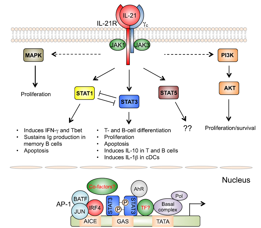
Figure 2: IL-21 and Its Signaling Pathway
STAT1 and STAT3 work together to enhance IL-21 expression, although they also exhibit opposing roles in the activation and cross-inhibition of signaling pathways. Studies have shown that in STAT3-deficient models, IL-21-mediated STAT1 phosphorylation is enhanced, suggesting that STAT3 may downregulate STAT1 phosphorylation.
In contrast, IL-2 and IL-15 strongly activate STAT5 but not STAT3. Data indicate that STAT3 is involved in multiple tumor signaling pathways. Research has demonstrated that inhibiting constitutive STAT3 activation suppresses the proliferation of ovarian and breast cancer cells, while sustained STAT3 activation promotes the proliferation of colorectal cancer cells and tumor growth.
IL-21 and Cells
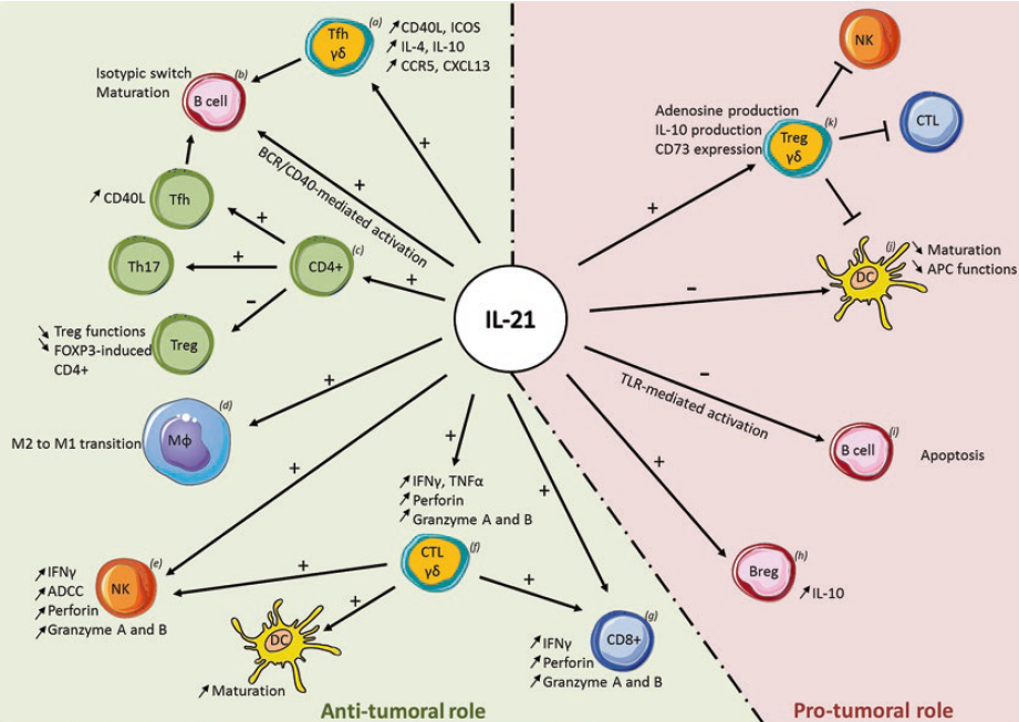
Figure 3: IL-21 and Different Cell Types
IL-21 and B Cells
The effects of IL-21 on B cells vary depending on the type and maturation state of the B cells. IL-21R expression differs among naïve B cells, memory B cells, and plasma cells. In the presence of IL-21, B cell activation (via B cell receptor or CD40 engagement) enhances their proliferation and differentiation into plasma cells, a process dependent on STAT3. Conversely, activation through Toll-like receptors can initiate B cell apoptosis in the presence of IL-21.
IL-21 is involved in B cell formation and germinal center (GC) function. GCs are structures formed by the massive proliferation of antigen-specific B cells after activation, primarily composed of B cells, follicular helper T cells (Tfh), follicular dendritic cells (FDCs), and macrophages. Studies have shown that the absence of IL-21 signaling severely impairs B cell responses to antigens and drastically reduces plasma cell formation in the spleen, bone, and bone marrow. Additionally, blocking IL-21 signaling reduces the expression of Bcl-6 in GC B cells.
IL-21 induces immunoglobulin class switching and effectively promotes plasmablasts to secrete IgM, IgG, IgA, and IgE. For naïve and memory B cells, IL-21 enhances immunoglobulin expression.
IL-21 antagonizes IgE expression. Research has shown that IL-21R-deficient mice exhibit IgE levels three times higher than those of control mice. IL-21 promotes the expression of Bmf (Bcl-2-modifying factor) in IgE-expressing B cells, which induces apoptosis, explaining why IgE levels remain high in the absence of IL-21.
IL-21 and NK Cells
Studies have shown that IL-21 enhances NK cell activity through the JAK/STAT and PI3K pathways across different species. In mouse models, IL-21 inhibits NK cell proliferation but induces NK cell activation, maturation, and the production of cytolytic factors (perforin and granzymes), thereby enhancing NK cell-mediated cytotoxicity. In humans, IL-21 also enhances NK cell cytotoxic activity. In vitro experiments demonstrate that IL-21 promotes NK cell survival and activation. Similar to observations in mice, IL-21 synergizes with IL-15 to enhance the production of cytolytic factors and IFN-γ.
IL-21 and T Cells
IL-21 inhibits the activation, differentiation, and survival of regulatory T cells (Tregs), thereby limiting their immunoregulatory functions. Tregs suppress T cell expression of IL-2 to exert immunosuppressive effects. Research has shown that IL-21R signaling in conventional T cells is necessary to counteract this suppression.
IL-21R signaling indirectly suppresses Treg-mediated immunosuppression by reducing IL-2 expression. IL-21 inhibits tumor-mediated FOXP3 expression in naïve CD4+ T cells, directly blocking their differentiation into Tregs.
Click on the product catalog numbers below to access detailed information on our official website.
This article is reviewed and published by the technical expert team of UA
Disclaimer: This article partially utilizes artificial intelligence assistance in its creation. If any content involves copyright or intellectual property issues, please let us know and we promise to verify and remove it as soon as possible.
Purchase recombinant protein, choose Nanjing UA-Bio
UA protein focuses on providing various protein reagents, raw materials, and services required for drug research and development, cell therapy, gene therapy, and basic scientific research, including drug target proteins, immune checkpoint proteins, cytokines, tool enzymes, customized protein expression, and full-length transmembrane protein development. Youai is committed to providing customers with high-quality products and professional services, and building a High-tech Biological Enterprise with International Competitiveness.

1.IL-21 sIgnaling in Immunity.
2.Tumor Microenvironment:The role of interleukins-Part A.
3.The role of IL-21 in Immunity and Cancer.






 Business license (three certificates in one)