




pH-Sensitive IgG Deep Red Labeling: Enabling Accurate Quantification and High Signal-to-Noise Ratio Imaging of Antibody Internalization


The pH-sensitive IgG labeling reagent (Deep Red) is a chemical tool for biological labeling, whose core characteristic lies in the significant alteration of fluorescence signal intensity with changes in environmental pH. This reagent is predominantly utilized for real-time, quantitative tracking of the internalization process of antibodies in live cells.
- Recent Advances
- Product Information
pH-Sensitive IgG Labeling Reagent (Deep Red) is a type of chemical tool for biological labeling, whose core characteristic is the significant change in fluorescence signal intensity with variations in environmental pH. This reagent is primarily used for the real-time and quantitative tracking of antibody internalization processes in live cells.
I. Basic Definition & Working Principle
This type of reagent contains two functional domains in its molecular structure:
-
Antibody-Binding Domain: This part can specifically and covalently bind to the Fc region of antibody molecules, enabling the labeling of IgG antibodies, typically without affecting the antibody's own antigen-binding capability.
-
pH-Sensitive Fluorescent Dye: This dye emits fluorescence in the deep red spectral region (excitation/emission wavelengths typically around 640/655 nm), and its fluorescence quantum yield is highly sensitive to environmental pH.
Its operation is based on a clear chemical principle:
-
Under neutral pH conditions (approximately pH 7.4), such as the extracellular environment or the cell membrane surface, the fluorophore of this dye is in a quenched state, producing only negligible fluorescence signal.
-
When the labeled antibody-antigen complex enters the cell via endocytosis, it is transported to acidic intracellular compartments, such as early endosomes, late endosomes, and lysosomes (pH range 4.5-6.0).
-
In the acidic environment, the chemical structure of the dye molecule changes, relieving the quenched state and causing a dramatic increase in fluorescence intensity, with an enhancement factor that can reach several hundred-fold.
II. Comparative Advantages Over Traditional Labeling Techniques
Compared to traditional fluorescent labeling methods, this technology offers the following distinct advantages:
-
Effectively Distinguishes Surface-Bound from Internalized Antibodies: Traditionally labeled antibodies produce strong fluorescence immediately outside the cell, resulting in high background signal that interferes with the detection of antibodies that have entered the cell. pH-sensitive labeling, through its "acidic environment activation"特性, confines the fluorescence signal to intracellular acidic compartments, thereby enabling specific imaging and quantification of internalized antibodies.
-
Excellent Optical Properties: The excitation and emission wavelengths of the Deep Red dye are in the far-red region. This spectral range offers two main advantages:
-
Low Autofluorescence: Autofluorescence from cells and tissues is very weak in this region, effectively reducing background noise.
-
Strong Tissue Penetrance: Far-red light penetrates biological tissues more effectively than visible light, making this technology more suitable for deep-tissue imaging and in vivo experiments in live animals.
-
-
Improved Accuracy for Quantitative Analysis: Since the turn-on of the fluorescence signal is directly linked to the intracellular acidic environment, the acquired data can more accurately reflect the kinetics and total amount of antibody endocytosis, providing a high signal-to-noise ratio detection method for quantitative studies.
III. Main Application Areas
This technology is primarily applied in the following research scenarios:
-
Antibody Internalization Studies: Real-time tracking of the uptake process of therapeutic antibodies or signaling antibodies by cells.
-
Antigen Presentation Research: Observing the trafficking and degradation fate of antigens bound by antibodies within cells.
-
Drug Development and Screening: Evaluating the internalization efficiency and intracellular trafficking pathways of antibody-based drugs (e.g., Antibody-Drug Conjugates ADCs).
-
Immunology and Cell Biology Research: Quantitative study of the internalization kinetics of cell surface receptors and their ligands.
IV. Conclusion
The pH-sensitive IgG labeling reagent (Deep Red), by coupling the fluorescence signal to the intracellular pH environment, provides a research tool capable of precisely distinguishing extracellular from intracellular events. Its optical properties in the far-red region, combined with low background and high signal-to-noise ratio detection capabilities, make it an advanced technological solution in the fields of live-cell imaging, quantitative analysis, and in vivo research.
Where to Buy pH-Sensitive IgG Labeling Reagents? Come to UA protein!
In antibody drug development, research on immune responses, and receptor-mediated internalization mechanisms, clearly distinguishing between cell surface binding and genuine internalization signals is a key challenge faced by researchers. High background fluorescence from traditional labeling methods severely interferes with data accuracy, making quantitative analysis difficult.
Leveraging its advanced bio-labeling technology platform, UA Biotech successfully launches two key products: pH-sensitive IgG labeling reagents (Deep Red) and pH-sensitive IgG labeling reagents (Red), offering you a dual-spectrum solution to completely overcome these challenges.
Overview of Core Product Advantages
-
Ultra-High Signal-to-Noise Ratio: Suppresses background at the source, yielding clean internalization signals.
-
Accurate Quantification: Provides reliable, quantifiable data for internalization kinetics studies.
-
Simple and Fast Procedure: The labeling process is straightforward and rapid, completable within 1 hour.
-
Excellent Compatibility: Suitable for various cell lines and adaptable to flow cytometry and high-throughput screening platforms.
-
Preserved Activity: Gentle labeling conditions ensure antibody activity and binding capacity remain unaffected.
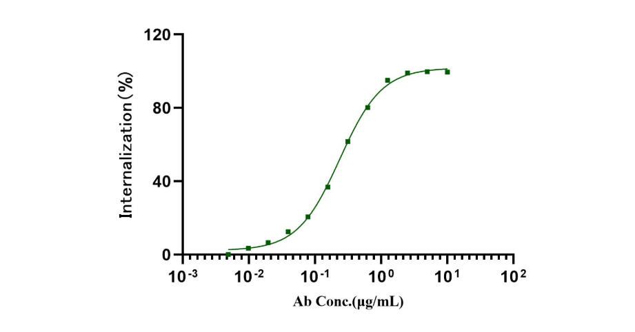 Fig1. The fluorescent intensity from antibody-probe protein complex internalization into cell is specific for B7-H3 on Hela cell.
This article is reviewed and published by the technical expert team of UA
Disclaimer: This article partially utilizes artificial intelligence assistance in its creation. If any content involves copyright or intellectual property issues, please let us know and we promise to verify and remove it as soon as possible.
Purchase recombinant protein, choose Nanjing UA-Bio
UA protein focuses on providing various protein reagents, raw materials, and services required for drug research and development, cell therapy, gene therapy, and basic scientific research, including drug target proteins, immune checkpoint proteins, cytokines, tool enzymes, customized protein expression, and full-length transmembrane protein development. Youai is committed to providing customers with high-quality products and professional services, and building a High-tech Biological Enterprise with International Competitiveness.







 Business license (three certificates in one)