




Choosing Between pH-Sensitive IgG Labeling Reagents (Red) and (Deep Red): A Comprehensive Guide


In cell biology and immunology research, tracking the internalization of antibodies in live cells is crucial. Traditional labeling methods often face challenges with high background fluorescence interference, while the emergence of pH-sensitive labeling technology has revolutionized the solution to this problem.
- Recent Advances
- Product Information
In cell biology and immunology research, tracking the internalization of antibodies in live cells is crucial. Traditional labeling methods often suffer from high background fluorescence interference, while pH-sensitive labeling technology has revolutionized the field by effectively addressing this issue. Among these, pH-sensitive IgG labeling reagents (Deep Red) and pH-sensitive IgG labeling reagents (Red) are two of the most commonly used star products. Although their names are similar, they have distinct differences in practical applications. This article provides an in-depth analysis of their core differences to help you make the best choice for your experiments.
I. Core Shared Principle: The Intelligently "Activated" Fluorescent Probe
First, understanding their shared working mechanism is the foundation for differentiation. Both reagents are fluorescent dyes covalently conjugated to IgG antibodies, with the most critical characteristic being their pH sensitivity:
Under Neutral or Alkaline pH (e.g., extracellular environment, pH ~7.4): The dye is in a quenched state, emitting almost no fluorescence. This means that non-internalized antibodies do not produce background signals, significantly improving the signal-to-noise ratio.
Under Acidic pH (e.g., inside endosomes/lysosomes, pH ~5.0-6.0): The fluorophore of the dye is protonated, undergoes a structural change, and is "activated," emitting strong fluorescence.
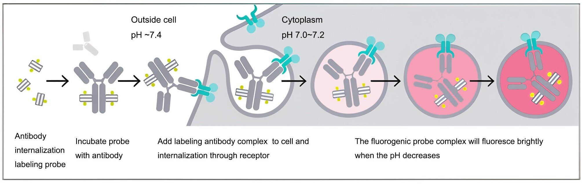
Therefore, when antibodies labeled with these reagents bind to cell surface antigens, only those engulfed by the cell and entering acidic organelles (endosomes, lysosomes) will fluoresce. Using fluorescence microscopy or flow cytometry, researchers can precisely observe and quantify the antibody internalization process.
II. Core Differences: Tissue Penetration and Autofluorescence
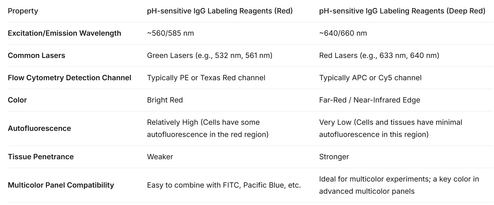
III. Antibody Incubation Protocols for pH-Sensitive IgG Labeling Reagents (Red) and (Deep Red)
A. pH-Sensitive IgG Labeling Reagents (Deep Red) - Example: UA BIOSCIENCE UA079026
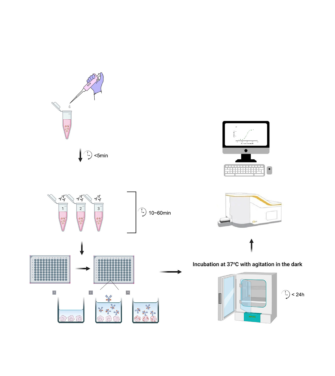
1.1 Incubation of UA079026 with Antibody
1.2 Reconstitute the lyophilized powder UA079026 according to the dissolution method using deionized water.
1.3 Prepare a sufficient volume of a 4X test antibody working solution in cell culture medium (four times the final test concentration, determined based on prior flow cytometry optimization). For example, 2 µg/ml might be a good starting concentration for the antibody, so the 4X working solution would be 8 µg/ml.
1.4 Prepare a sufficient volume of a 4X UA079026 working solution in cell culture medium. Use a molar ratio of Antibody:UA079026 = 1:2.
1.5 Mix the 4X test antibody working solution and the 4X UA079026 working solution in a 1:1 volume ratio. Incubate at room temperature protected from light for 15 min - 1 hour to obtain the Ab-UA079026 complex 2X working solution.
2. Incubation of Ab-UA079026 Complex with Cells
2.1 Cell Preparation: Harvest and wash cells. Adjust cell concentration using complete medium. For suspension cells: 1-2×10^5 cells/mL; for adherent cells: 0.5-1×10^5 cells/mL. Add 100 µL of cell suspension per well in a 96-well plate.
2.2 Incubation: Add 100 µL of the Ab-UA079026 complex 2X working solution to each well. Culture in a 37°C, 5% CO2 incubator.
3. Flow Cytometry Detection
After culturing for 18-24 hours (adjustable based on the actual internalization kinetics of the antibody), harvest the cells and analyze antibody internalization by flow cytometry using the Cy5 channel.
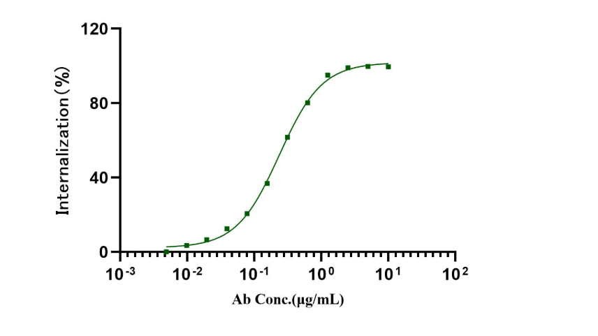
Fig1. The fluorescent intensity from antibody-probe protein complex internalization into cell is specific for B7-H3 on Hela cell.
pH-sensitive lgG labeling reagents (Deep Red)_UA079026_优爱(UA BIOSCIENCE)官网
B. pH-Sensitive IgG Labeling Reagents (Red) - Example: UA BIOSCIENCE UA070127
Incubation of UA070127 with Antibody
1.1 Reconstitute the lyophilized powder UA070127 according to the dissolution method using deionized water.
1.2 Prepare a sufficient volume of a 4X test antibody working solution in cell culture medium (four times the final test concentration, determined based on prior flow cytometry optimization). For example, 2 µg/ml might be a good starting concentration for the antibody, so the 4X working solution would be 8 µg/ml.
1.3 Prepare a sufficient volume of a 4X UA070127 working solution in cell culture medium. Use a molar ratio of Antibody:UA070127 = 1:2.
1.4 Mix the 4X test antibody working solution and the 4X UA070127 working solution in a 1:1 volume ratio. Incubate at room temperature protected from light for 15 min - 1 hour to obtain the Ab-UA070127 complex 2X working solution.
2. Incubation of Ab-UA070127 Complex with Cells
2.1 Cell Preparation: Harvest and wash cells. Adjust cell concentration using complete medium. For suspension cells: 1-2×10^5 cells/mL; for adherent cells: 0.5-1×10^5 cells/mL. Add 100 µL of cell suspension per well in a 96-well plate.
2.2 Incubation: Add 100 µL of the Ab-UA070127 complex 2X working solution to each well. Culture in a 37°C, 5% CO2 incubator.
3. Flow Cytometry Detection
After culturing for 18-24 hours (adjustable based on the actual internalization kinetics of the antibody), harvest the cells and analyze antibody internalization by flow cytometry using the PE channel.
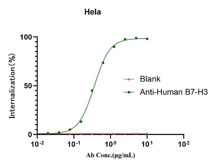
Fig1. The fluorescent intensity from antibody-probe protein complex internalization into cell is specific for B7-H3 on Hela cell.
pH-sensitive lgG labeling reagents (Red)_UA070127_优爱(UA BIOSCIENCE)官网
Where to Buy pH-Sensitive IgG Labeling Reagents?
UAbio leverages its advanced biomarking technology platform to successfully offer two flagship products: pH-sensitive IgG labeling reagents (Deep Red) and pH-sensitive IgG labeling reagents (Red), providing you with a dual-spectrum solution to thoroughly overcome the challenges mentioned above.
This article is reviewed and published by the technical expert team of UA
Disclaimer: This article partially utilizes artificial intelligence assistance in its creation. If any content involves copyright or intellectual property issues, please let us know and we promise to verify and remove it as soon as possible.
Purchase recombinant protein, choose Nanjing UA-Bio
UA protein focuses on providing various protein reagents, raw materials, and services required for drug research and development, cell therapy, gene therapy, and basic scientific research, including drug target proteins, immune checkpoint proteins, cytokines, tool enzymes, customized protein expression, and full-length transmembrane protein development. Youai is committed to providing customers with high-quality products and professional services, and building a High-tech Biological Enterprise with International Competitiveness.







 Business license (three certificates in one)